Recent advancements in the realm of neonatal health research have illuminated the intricate relationship between brain development and metabolic processes in preterm infants. A compelling study conducted by Xu and colleagues has employed magnetic resonance imaging (MRI) techniques to evaluate both glutamate levels and morphometric changes in the hippocampus of low-birth-weight preterm infants at term-equivalent age. This groundbreaking research showcases advancements in diagnostic imaging that allow for a more profound understanding of the biological complexities that affect infants born before their due date.
The hippocampus, a pivotal region of the brain associated with memory formation and emotional regulation, undergoes significant developmental changes shortly after birth. Infants born preterm, particularly those with low birth weight, face a higher risk of neurodevelopmental challenges due to underdeveloped neural pathways and metabolic dysregulation. The impact of a low birth weight on the Newborn’s hippocampal structure and resulting cognitive capabilities underscores the necessity for effective monitoring and intervention strategies.
Glutamate, the brain’s primary excitatory neurotransmitter, plays an essential role in synaptic plasticity and neurodevelopment. In the context of preterm infants, altered glutamate signaling may contribute to neurodevelopmental disorders and cognitive impairments. The assessment conducted by Xu et al. involves advanced MRI techniques to quantify hippocampal glutamate levels, providing critical insight into the neurochemical environment supporting neural growth and connectivity. This method offers a non-invasive window into the brain’s metabolic state, allowing clinicians and researchers to correlate findings with cognitive outcomes.
As we delve deeper into the implications of the research, it becomes evident that the combination of imaging glutamate levels and morphometric analysis reveals the nuances of brain development in preterm infants. By employing high-resolution MRI scans, the researchers could not only measure glutamate concentrations but also assess variations in hippocampal volume, surface area, and shape. These morphometric changes offer insights into how early brain development may influence long-term neurocognitive abilities, establishing a tangible connection between physiology and psychological health.
One particularly interesting finding from Xu’s study is the specific alterations in hippocampal morphology observed in preterm infants with low birth weight. The research has indicated that these infants often display a reduction in hippocampal volume compared to their term-born counterparts. This reduction might signal impaired neurogenesis and increased vulnerability to behavioral and cognitive deficits as they age. By illuminating these early physiological changes, the study encourages ongoing exploration into targeted neuroprotective strategies that could foster cognitive development in at-risk populations.
The methodology employed within this study showcases the remarkable capabilities of modern imaging technologies. MRI has progressed significantly, allowing researchers to explore the structure and function of the brain in unprecedented detail. This non-invasive approach avoids the complications and risks associated with other imaging modalities, such as X-rays or CT scans, thereby providing a safe option for examining delicate neural structures in vulnerable populations, such as preterm infants.
Furthermore, the implications of assessing glutamate levels through MRI extend beyond mere academic inquiry. The potential for early intervention based on identified neurochemical imbalances presents new avenues for therapeutic strategies aimed at mitigating the impact of preterm birth-related neurodevelopmental issues. If clinicians can set benchmarks for neural health based on glutamate assessment, it could lead to tailored interventions that optimize cognitive outcomes over time.
In contemplating the broader ramifications of this research, it is crucial to consider socioeconomic factors and how they intersect with neonatal health outcomes. Low birth weight commonly aligns with elements such as maternal health, access to prenatal care, and environmental stressors, all of which influence brain development in infants. The findings from Xu et al. not only contribute to a better understanding of neurodevelopmental processes but also serve as a reminder of the multifaceted challenges faced by families and healthcare providers working with preterm infants.
It’s imperative for ongoing research to identify the interconnectedness of nutrition, genetics, and environmental influences, creating a comprehensive framework for understanding how to best support cognitive outcomes in low-birth-weight infants. The evaluations presented in this study are just the tip of the iceberg in exploring the myriad factors affecting infant health, thereby fostering a more holistic approach to neonatal care.
In summary, the research presented by Xu and colleagues embodies an essential leap forward in understanding how preterm birth and low birth weight impact brain development. By utilizing sophisticated MRI techniques to monitor glutamate levels and hippocampal changes, the study shines a light on the intricate interplay between nutrition, cognitive health, and neurological development. Such insights are critical, not only for the current generation of preterm infants but also for informing future healthcare strategies aimed at improving neonatal outcomes.
Ultimately, the call for research and clinical attention to neurodevelopment in preterm infants has never been more urgent. The wealth of information garnered from studies such as these can significantly shape our understanding and response to the complex challenges of early brain development. As scientists and healthcare providers collaborate to translate these findings into practical intervention strategies, the future of neonatal care holds great promise for enhanced cognitive and emotional outcomes in populations that have previously grappled with substantial risks.
Subject of Research: Assessment of hippocampal glutamate levels and morphometric changes in preterm infants with low birth weight.
Article Title: Magnetic resonance imaging-based assessment of hippocampal glutamate and morphometric changes in preterm infants at term-equivalent age with low birth weight.
Article References: Xu, L., Gong, H., Ren, Q. et al. Magnetic resonance imaging-based assessment of hippocampal glutamate and morphometric changes in preterm infants at term-equivalent age with low birth weight. Pediatr Radiol (2025). https://doi.org/10.1007/s00247-025-06435-8
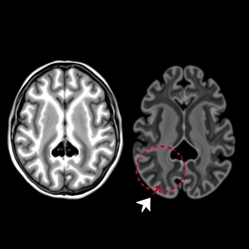
Image Credits: AI Generated
DOI: https://doi.org/10.1007/s00247-025-06435-8
Keywords: Neonatal health, MRI, hippocampus, glutamate, preterm infants, low birth weight, neurodevelopment.
