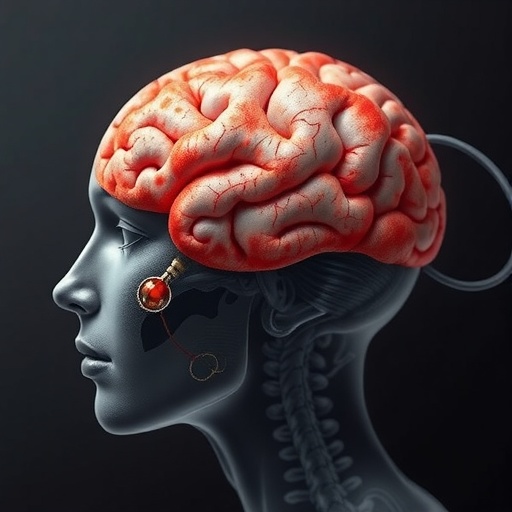
In a groundbreaking study poised to reshape our understanding of brain plasticity, researchers at the University of Pittsburgh School of Medicine and Cambridge University have discovered that the brain’s somatosensory map remains strikingly stable even after the amputation of a limb. Published in the prestigious journal Nature Neuroscience, this research overturns decades of neuroscientific dogma that assumed dramatic cortical reorganization in response to limb loss. The findings, which challenge entrenched paradigms, hold promising implications for refining treatments of phantom limb pain and advancing brain-computer interface technologies aimed at restoring sensation and control over prosthetic limbs.
For over fifty years, the prevailing belief in neuroscience has held that the brain’s somatosensory cortex undergoes significant and rapid remapping after the physical loss of a body part. This cortical reorganization hypothesis proposed that neighboring brain regions would expand into the now-deafferented territory that previously represented the amputated limb. For instance, following the loss of a hand, it was thought that adjacent cortical areas—such as those corresponding to the lips or face—would ‘invade’ and repurpose that territory. This theory, though widely accepted, never fully reconciled with patient-reported experiences of vivid, stable sensations emanating from missing limbs, often manifesting as the phenomena collectively termed “phantom limb sensations”.
The new study, led by Dr. Tamar Makin of Cambridge University and Dr. Hunter Schone of the University of Pittsburgh’s Rehab Neural Engineering Labs, is the first to provide direct longitudinal evidence of sensorimotor cortical stability before and after hand amputation. By leveraging cutting-edge functional magnetic resonance imaging (fMRI), the research team examined cortical activity in three individuals scheduled for elective hand amputation. Importantly, imaging sessions occurred both prior to surgery and at multiple points afterward — three, six, and in some cases, eighteen months to five years post-amputation — allowing an unprecedented dynamic assessment of brain remapping processes over time.
During the fMRI sessions, participants were instructed to move or attempt to move their fingers and to purse their lips, actions designed to activate discrete, topographically defined regions within the primary somatosensory cortex. Contrary to conventional expectations, the cortical representation of the missing hand remained largely preserved, with patterns of brain activation mirroring those observed before limb loss. Moreover, the facial area of the somatosensory cortex, specifically the lips region, showed no evidence of encroaching upon or taking over the hand representation zone, conclusively refuting the long-held belief in extensive post-amputation cortical reorganization.
This remarkable persistence of the ‘body map’ may be rooted in the underlying architecture of the somatosensory cortex, where overlapping and distributed neural networks encode multisensory inputs. The researchers propose that the assumption of ‘invasion’ by neighboring cortical territories was a misinterpretation arising from the coarse spatial resolution of earlier imaging techniques and methodologies that failed to account for the intrinsic complexity of sensorimotor representations. Instead of a simplistic rearrangement, the brain maintains an enduring template of the body, retaining latent circuitry for the missing limb that remains functionally accessible.
The implications of this discovery extend well beyond academic debates about neuroplasticity. Phantom limb pain, a debilitating condition affecting a large proportion of amputees, has long been attributed to maladaptive cortical reorganization. Therapeutic attempts aimed at ‘correcting’ these supposed reconfigurations, however, have historically been met with limited success. The new findings redirect focus toward the peripheral nervous system and the role of aberrant afferent signaling in generating phantom sensations and pain. Reconstructive surgical techniques that re-route residual nerves to newly innervated muscle or skin have demonstrated promising outcomes, including significant pain relief in study participants who underwent such procedures following amputation.
Furthermore, this research carries profound significance for the future of neuroprosthetics and brain-computer interfaces (BCIs). These cutting-edge technologies depend on decoding precise neural activity patterns to restore sensation and motor control in paralyzed or amputated limbs. The demonstrated stability of cortical limb representations suggests that BCI systems can reliably interface with existing neural circuitry over extended periods, without concern for dynamic remapping—a crucial step toward developing more sophisticated prosthetic devices that convey rich sensory feedback and nuanced motor commands.
Dr. Schone emphasized the transformative potential of these insights: “Knowing that the somatosensory body map is stable enables us to push the envelope in neural engineering. We can now target increasingly finer scales within the hand area—the ability to distinguish activation patterns from a fingertip versus the base of a finger—and work toward restoring complex sensations like texture, shape, and temperature through brain-computer interfaces.”
The paradigm-shifting nature of this study also encourages a reexamination of previous neuroimaging studies of amputation and cortical plasticity. The authors caution that methodological limitations and oversimplified interpretations may have led to erroneous conclusions about brain reorganization in human and animal models alike. They advocate for future research employing higher-resolution imaging, refined analytic techniques, and longitudinal designs to unravel the nuances of sensorimotor cortical function in health and disease.
Moreover, this study offers a hopeful narrative for amputees experiencing phantom sensations. Rather than depicting the brain as a malleable yet unstable organ constantly reshaped by sensory loss, it depicts a resilient neural substrate ‘waiting to reconnect,’ preserving the essence of the missing hand. This intrinsic fidelity implies that therapeutic interventions may harness these latent pathways, promoting more effective sensory restoration and potentially enhancing functional recovery.
In concert with experts from the National Institutes of Health and other institutions, the collaborative team envisions ongoing studies to extend these findings to larger cohorts and investigate the molecular and cellular mechanisms underpinning cortical map stability. This integrative inquiry is essential for translating fundamental neuroscience discoveries into clinical innovations to improve quality of life for individuals with limb loss and related neurological conditions.
As the neuroplasticity paradigm shifts, the scientific community must grapple with the broader implications of a brain that resists wholesale reorganization despite drastic physical alterations. This revelation compels not only a reassessment of brain adaptability but also invigorates optimism for neurotechnological advancements and rehabilitative medicine. Ultimately, the legacy of this research lies in its capacity to unify cutting-edge neuroscience, clinical insight, and engineering ingenuity toward restoring the intimate connection between mind and body, even in the face of profound loss.
Subject of Research: Neuroscience – Brain Plasticity and Somatosensory Cortex Stability Following Limb Amputation
Article Title: Brain’s Somatosensory Map Remains Stable After Hand Amputation, Challenging Long-Held Views on Cortical Plasticity
News Publication Date: August 21, 2025
References: Schone et al., Nature Neuroscience, 2025
Image Credits: Schone et al., Nature Neuroscience, 2025
Keywords: Brain, Nervous system, Neuroscience, Clinical neuroscience, Neuroimaging, Neurophysiology, Human brain, Motor control, Neural pathways, Neuroplasticity, Cortical maps, Sensory systems, Pain, Chronic pain, Neuropathic pain


