


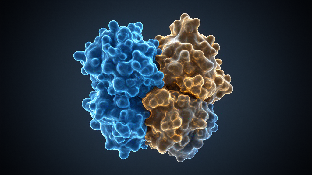
Published in Applied Microscopy, the study presents a novel multi-stain approach applied to the pyruvate dehydrogenase E2 (PDH E2) complex, a large macromolecular assembly from Bacillus stearothermophilus. By integrating data from uranyl acetate, ammonium phosphotungstate, and ammonium molybdate, the team achieved a resolution of approximately 21.7 Å, a notable improvement over the 27.2 to 31.4 Å range typically observed when using these stains individually. This technique offers a practical method for refining low-resolution structural models, bridging the gap between initial sample characterization and high-resolution structural biology studies.
Negative staining electron microscopy has long been a cornerstone technique in structural biology, valued for its rapid and accessible method for visualizing protein structures. The process involves embedding biological specimens in a thin layer of heavy metal salts, which creates a contrast-enhancing electron-dense background that outlines the specimen’s topography. Despite its widespread utility, the method has inherent limitations, including potential staining artifacts and the risk of sample flattening. These constraints often result in incomplete structural information, particularly for large, multi-subunit protein complexes that possess intricate architectures with diverse structural elements.
The choice of heavy metal stain significantly influences image quality and interpretation in negative staining electron microscopy. Commonly used stains such as uranyl acetate (UA), ammonium phosphotungstate (PTA), and ammonium molybdate (AM) each offer unique advantages due to their distinct physicochemical properties. UA is positively charged and binds to negatively charged regions on the protein surface, providing high contrast and fine grain that excel in overall shape visualization. In contrast, PTA and AM are negatively charged and highlight positively charged areas, offering different perspectives on the protein’s surface topology.
The PDH E2 complex serves as an ideal model system for exploring these multi-stain approaches due to its complex architecture. The complex forms a roughly spherical structure with an approximate diameter of 25–30 nm, composed of 20 trimeric units arranged in an icosahedral symmetry. Surface charge analysis revealed a striking electrostatic landscape, with strong positive charges in the regions between trimers and alternating patches of positive and negative charges on the trimer surfaces. This heterogeneous charge distribution likely plays a key role in stabilizing interactions between adjacent trimers and facilitating interactions with various substrates.
In the study, purified PDH E2 complex samples were subjected to negative staining using the three different heavy metal stains. For each staining method, 5 µL of the complex was applied to a glow-discharged carbon-coated copper grid and allowed to adsorb for 30 seconds. The grids were then stained with 1% solutions of UA, PTA, or AM, followed by blotting and air-drying. Micrographs were collected on a Tecnai 10 transmission electron microscope operated at 100 kV, with 300 micrographs recorded for each staining condition. This systematic application allowed the researchers to compare the structural features revealed by each stain.
Each staining method revealed distinct structural features of the PDH E2 complex, providing complementary information about its architecture. UA staining provided high contrast and fine structural detail, clearly delineating the overall shape of the complex. PTA staining resulted in slightly lower contrast but offered improved preservation of the complex’s native structure, particularly at the periphery. AM staining produced images with softer contrast but excellent structural preservation, highlighting the complex’s symmetrical nature and maintaining the integrity of delicate structural features.
The analysis of 2D class averages showed distinct differences in the apparent size of the PDH E2 complex under different staining conditions. UA staining resulted in particles with an average long axis of 264.0 ± 3.6 Å, while PTA staining produced larger particle images with an average long axis of 284.8 ± 8.8 Å. AM yielded the smallest particle images, with an average long axis of 253.6 ± 7.7 Å. These variations can be explained by the distinct interactions of each staining agent with the protein surface. UA closely approximates the true dimensions from the crystal structure, while PTA creates a more substantial stain envelope due to its attraction to positively charged patches, and AM penetrates into the complex’s cavities, minimizing the external stain layer thickness.
When all staining methods were combined into a single dataset, two distinct size populations emerged, indicating effective integration of particles from different staining methods. The combined dataset achieved a markedly improved resolution of 21.7 Å, compared to the 31.4 Å resolution from UA, 30.3 Å from PTA, and 27.2 Å from AM. This enhancement facilitated a clearer visualization of the complex’s icosahedral symmetry and allowed for a more precise determination of the overall shape and domain organization. The multi-stain approach revealed complementary structural information, with each stain highlighting different aspects of the protein complex.
Local resolution analysis using ResMap further demonstrated the benefits of the multi-stain approach. The combined reconstruction exhibited predominantly cyan coloration, indicating higher overall resolution of 10–12 Å throughout the complex, compared to the single-stain reconstructions which predominantly showed magenta coloration corresponding to 14–18 Å resolution. This improvement suggests that integrating multiple stains averages out artifacts and inconsistencies introduced by individual stains, yielding a more uniform and detailed reconstruction. The local resolution maps provided valuable information about the structural details preserved by each staining method, with areas of higher resolution particularly visible in the inner cavities and along structural interfaces.
It is crucial to contextualize the resolution values reported within the limitations of negative staining electron microscopy. Unlike cryo-EM, where resolution serves as a robust indicator of structural detail, the concept of resolution in negative staining reconstructions has different implications. Negative staining relies on heavy metal salts to outline the protein’s surface, producing 2D projections rather than capturing internal structural details. Consequently, the resolutions achieved are typical for this technique and do not approach the near-atomic precision attainable with cryo-EM. The improvement from 31.4 Å to 21.7 Å is notable within the scope of negative staining, reflecting an enhancement in surface detail rather than a fundamental leap in structural resolution.
The success of this multi-stain approach stems from the complementary nature of the information provided by each stain. By combining these datasets, the researchers effectively mitigated limitations inherent to single-stain methods, such as incomplete visualization due to selective stain binding. The method proved particularly effective for the PDH E2 complex, given its heterogeneous surface charge distribution. However, the effectiveness of the multi-stain strategy may vary depending on the specific surface properties and structural stability of the protein complex under study. For complexes lacking the charge diversity or rigidity of large protein assemblies, benefits might be less pronounced.
Despite these constraints, the multi-stain approach offers a practical and accessible means to obtain a more accurate initial model of complex protein structures. For the PDH E2 complex, it has provided valuable insights into its overall shape and organization, serving as a foundation for subsequent high-resolution investigations. While not a replacement for advanced techniques like cryo-EM, this method represents a promising option for structural biologists seeking to refine low-resolution models in the early stages of analysis, particularly for challenging macromolecular assemblies.
Looking forward, this multi-stain negative staining approach could be especially valuable for analyzing diverse structural questions in macromolecular biology. It could be applied to study conformational changes in large protein complexes under different functional states, characterize the architecture of membrane protein assemblies, and provide foundation models for integrative structural biology approaches. By extracting more comprehensive low-resolution structural information, this approach bridges the critical gap between initial sample characterization and detailed structural analysis, potentially accelerating our understanding of complex macromolecular machines that underpin cellular functions.
Subject of Research: Multi-stain negative staining electron microscopy for protein complex structural analysis
Article Title: Technical approaches of 3D reconstruction from protein complex using the mixture of differently stained images: providing suggestive evidence for improving its resolution
Article References: Park, Y. H., Jeong, M. S., Song, G. S., gwonbaek, T., Kim, Y. K., & Jung, H. S. (2025). Technical approaches of 3D reconstruction from protein complex using the mixture of differently stained images: providing suggestive evidence for improving its resolution. Applied Microscopy, 55(1), Article 6. https://doi.org/10.1186/s42649-025-00111-9
Image Credits: AI Generated
DOI: 10.1186/s42649-025-00111-9
Keywords: electron microscopy, protein structure, negative staining, structural biology, pyruvate dehydrogenase, resolution enhancement, heavy metal stains, icosahedral symmetry, Technical, approaches, reconstruction, protein
Cite Scienmag News
Jason Bradley. (November 12, 2026). Multi-Stain Electron Microscopy Improves Protein Complex Resolution. Scienmag. https://scienmag.com/multi-stain-electron-microscopy-improves-protein-complex-resolution/
Jason Bradley. "Multi-Stain Electron Microscopy Improves Protein Complex Resolution." Scienmag, 12 November 2026, https://scienmag.com/multi-stain-electron-microscopy-improves-protein-complex-resolution/. Accessed 12 November 2026.
Jason Bradley. "Multi-Stain Electron Microscopy Improves Protein Complex Resolution." Scienmag. November 12, 2026. https://scienmag.com/multi-stain-electron-microscopy-improves-protein-complex-resolution/




