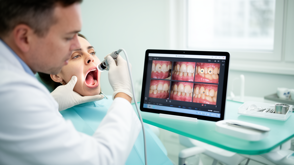
Deep learning models can sort close-up photographs of teeth into categories of enamel decay with striking accuracy, but a new study argues that the field has been measuring those claims badly. In a systematic benchmark published in Discover Imaging, researchers trained five convolutional neural network architectures on the same publicly available set of 2,000 intraoral color photographs and evaluated them under a single, tightly controlled protocol, finding that a compact pretrained network called EfficientNet-B0 delivered the best numerical results while a hand-built custom network trailed far behind.
The study, led by Komal Vyas, Devesh Nawgaje and Ashwini Deshmukh, used the Caries_Spectra dataset, a freely available collection of 2,000 RGB intraoral images gathered from clinics in Rajshahi and Dhaka, Bangladesh, between 2021 and 2022. The images fall into three diagnostic classes: 800 showing advanced enamel caries, 800 showing early-stage enamel caries, and 400 showing no enamel caries. Rather than relying on a single train-test split, the team ran stratified five-fold cross-validation twice with different random seeds, producing ten independent evaluation runs for every architecture. Because the public dataset contains no patient identifiers, patient-wise splitting was impossible, so the authors instead guaranteed that no image appeared in both the training and validation subsets of any fold.
All five models, ResNet18, MobileNetV2, EfficientNet-B0, DenseNet121 and a custom convolutional network, went through identical preprocessing. Every image was resized to 224 by 224 pixels and normalized with a mean and standard deviation of 0.5 per channel. Training images alone received augmentation in the form of random horizontal flips, rotations within 15 degrees, and brightness and contrast shifts of 0.2, while validation folds saw only deterministic resizing and normalization. The four pretrained networks started from ImageNet weights with their final layers replaced to output the three classes; the custom CNN, built from three convolutional blocks with batch normalization, ReLU activation and max pooling, was trained from scratch as a baseline.
Training was carried out entirely on a CPU-based Linux system with 24 cores and roughly 126 gigabytes of RAM, with no GPU acceleration, using PyTorch, the Adam optimizer at a learning rate of 0.0001, a batch size of 32, cross-entropy loss, and early stopping that halted training after seven epochs without meaningful improvement in validation loss. The authors report that this uniform setup was chosen to eliminate differences caused by training procedure rather than architecture, a common confound in dental AI studies where heterogeneous datasets, validation schemes and metrics make published results difficult to compare.
The headline result: EfficientNet-B0 achieved a mean accuracy of 98.18 percent with a standard deviation of 0.82, and a 95 percent confidence interval of 97.59 to 98.76 percent, along with an F1-score, precision and recall each at 0.985. DenseNet121 matched that mean accuracy with a confidence interval of 97.66 to 98.69 percent. ResNet18 reached 97.83 percent accuracy and MobileNetV2 97.65 percent, while the custom CNN managed only 91.28 percent with markedly higher variability, its confidence interval spanning 89.63 to 92.92 percent. Statistical stability tests found no significant differences between repeated runs of the same architecture, supporting the reproducibility of the training process.
When the architectures were compared against each other, the Friedman test showed highly significant differences in accuracy, F1-score, precision and recall, with chi-square values around 25 and p-values below 0.001. Post hoc Wilcoxon signed-rank tests with Bonferroni correction revealed that every pretrained model significantly outperformed the custom CNN, with an adjusted p-value of 0.0195 for each comparison, but that no statistically significant difference separated the pretrained networks from one another. In other words, EfficientNet-B0’s numerical lead, however small, does not prove architectural superiority; the authors explicitly caution against reading small accuracy gaps as evidence that one network is better than another.
Discrimination analysis told a similar story. Macro-averaged ROC-AUC values, computed per class and averaged across all ten runs, ranged from 0.9964 to 0.9979 for the pretrained models, with MobileNetV2 nominally highest at 0.9979 plus or minus 0.0015, followed closely by EfficientNet-B0 and ResNet18, while the custom CNN reached 0.9850. Class-wise analysis showed that all pretrained networks were best at recognizing the no-caries category, and that the hardest distinction was between advanced and early-stage enamel caries. Confusion matrices confirmed that nearly all misclassifications occurred between those two adjacent disease stages, while healthy enamel was reliably identified with few false positives, suggesting the real challenge for automated caries classification lies in staging lesions, not in distinguishing diseased from sound tissue.
Computational profiling added a practical dimension. The custom CNN carried the most trainable parameters, 25.78 million, yet required the fewest floating-point operations at 0.19 billion, a reminder that parameter count alone does not determine computational cost. DenseNet121 was the lightest in parameters at 6.96 million but the heaviest in computation, needing 2.88 billion FLOPs and 40.3 minutes of training per fold. MobileNetV2 trained fastest at 16.7 minutes per fold, EfficientNet-B0 close behind at 17.1 minutes with only 4.01 million parameters, and ResNet18 took 25.0 minutes. Convergence behavior tracked these differences: the pretrained networks stabilized after roughly 20 to 23 epochs, while the custom CNN needed about 35. The authors highlight EfficientNet-B0’s combination of top accuracy and moderate cost as promising for resource-limited clinical settings.
To peer inside the models, the team applied Grad-CAM, a gradient-weighted saliency technique that highlights image regions driving each prediction. Visualizations were generated only for correctly classified images with softmax confidence above 0.90, yielding 376 images for EfficientNet-B0, 371 for DenseNet121, 367 for MobileNetV2, 363 for ResNet18 and 188 for the custom CNN. Qualitatively, the better-performing pretrained networks produced activation maps concentrated on visually recognizable areas of enamel demineralization, whereas MobileNetV2 and the custom CNN often spread activation broadly beyond lesion regions. The authors stress that these maps are qualitative only: because Caries_Spectra lacks pixel-level lesion annotations, no quantitative localization check was possible, and saliency maps in medical imaging are known to sometimes latch onto lighting artifacts or structural priors rather than pathology.
The study’s authors position the work as one of the first reproducible benchmarks on this dataset, and they situate it against prior reports of 88.95 to 99.25 percent accuracy from studies using specialized architectures, multimodal inputs or different validation schemes, comparisons they describe as numerically non-comparable because of methodological differences. The limitations they acknowledge are substantial: evaluation on a single dataset with no external validation, no information about who labeled the images or how, possible correlations between images from the same patient or tooth, and a scope restricted to three enamel categories on RGB photographs, excluding dentinal or root caries and other oral conditions. They frame the models strictly as decision-support tools for clinicians rather than replacements for professional examination, and they call for external multicenter validation, expert-annotated lesion regions, uncertainty estimation and prospective clinical testing before any real-world deployment.
Subject of Research: Benchmarking deep learning models for three-class enamel caries classification on intraoral photographs with cross-validation and Grad-CAM interpretability
Article Title: Performance comparison of deep learning models for automated dental caries classification with Grad-CAM visualization
Article References: Vyas, K., Nawgaje, D., & Deshmukh, A. (2026). Performance comparison of deep learning models for automated dental caries classification with Grad-CAM visualization. Discover Imaging, 3(1), Article 11. https://doi.org/10.1007/s44352-026-00032-7
Image Credits: AI Generated
DOI: 10.1007/s44352-026-00032-7
Keywords: deep learning, dental caries, enamel caries, Caries_Spectra dataset, EfficientNet-B0, convolutional neural networks, cross-validation, Grad-CAM, intraoral images, transfer learning, medical imaging, model benchmarking
Cite Scienmag News
Ophelia Keating. (October 27, 2026). Head-to-head benchmark ranks EfficientNet-B0 top for spotting enamel caries in intraoral photos. Scienmag. https://scienmag.com/head-to-head-benchmark-ranks-efficientnet-b0-top-for-spotting-enamel-caries-in-intraoral-photos/
Ophelia Keating. "Head-to-head benchmark ranks EfficientNet-B0 top for spotting enamel caries in intraoral photos." Scienmag, 27 October 2026, https://scienmag.com/head-to-head-benchmark-ranks-efficientnet-b0-top-for-spotting-enamel-caries-in-intraoral-photos/. Accessed 28 October 2026.
Ophelia Keating. "Head-to-head benchmark ranks EfficientNet-B0 top for spotting enamel caries in intraoral photos." Scienmag. October 27, 2026. https://scienmag.com/head-to-head-benchmark-ranks-efficientnet-b0-top-for-spotting-enamel-caries-in-intraoral-photos/