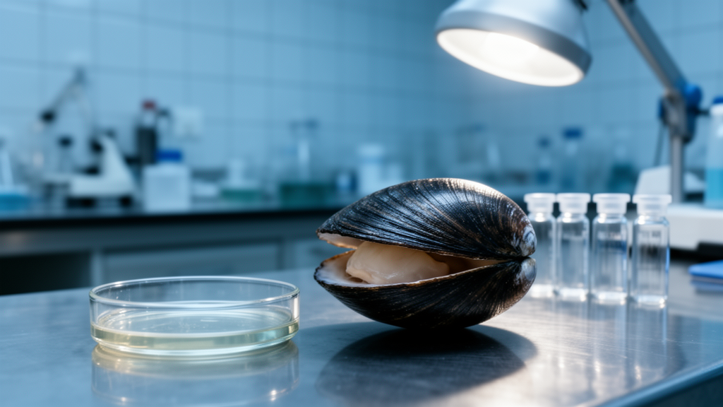
The ocean quahog Arctica islandica can live for more than five centuries, making it the longest-lived non-colonial animal known. Why some bivalve species survive so much longer than close relatives with similar body plans has long puzzled biologists, and mitochondria have repeatedly surfaced as a leading suspect. A new comparative study adds a missing piece to that puzzle: a systemic map of how metabolism itself differs across the lifespan gradient. By quantifying 50 metabolites in two tissues of five bivalve species spanning maximum reported longevities from 28 to 507 years, researchers found that longevity is written into the metabolic profile of the animals, with the tricarboxylic acid (TCA) cycle and amino acid metabolism emerging as the central signatures.
The team compared five species collected in eastern Canada: Mya arenaria (28 years), Spisula solidissima (37 years), Mactromeris polynyma (92 years), Mercenaria mercenaria (106 years) and Arctica islandica (507 years). All animals were mature adults, held for at least six months under standardized seasonal conditions at a marine research station, and sampled from gill and mantle tissue after a brief incubation approximating resting physiology. Sample sizes were modest, ranging from four to eight individuals per species, with the same individuals used for both tissues. The targeted panel covered amino acids, TCA cycle and glycolytic intermediates, nucleotides, and anaerobic metabolites such as lactate, alanine, strombine and octopine, quantified by HPLC coupled to triple quadrupole mass spectrometry and expressed per milligram of dry tissue.
The first striking result was that species identity and tissue type, but not body size, shaped the global metabolomic profile. Multivariate testing found significant effects of species, tissue and their interaction on metabolomic patterns, while body size showed no detectable influence. In unsupervised principal component analysis of gill tissue, the first principal component, accounting for 23 percent of variance, aligned with maximum reported longevity, forming a continuous gradient across species. That axis was driven mainly by essential amino acids, including branched-chain amino acids, threonine and phenylalanine, along with the non-essential amino acids serine, glycine and alanine. In mantle tissue, longevity-related variation spread across the second and third components, the latter dominated by the TCA intermediates fumarate and malate. A supervised model confirmed the same species-level separation and lifespan gradient.
Because the ocean quahog sits at such an extreme end of the lifespan range, the authors also repeated their key regression analyses without it. Even with the reduced lifespan range and statistical power, the principal metabolic trends remained evident, although support for several TCA-cycle associations weakened. Conversely, an exploratory analysis excluding the shortest-lived species, M. arenaria, which faces recurrent hypoxia and osmotic stress in its intertidal habitat, strengthened several longevity-associated trends. The overall metabolic signature was therefore not solely an artifact of either extreme endpoint, though the authors stress that these analyses are hypothesis-generating rather than definitive.
The clearest longevity-linked pattern involved a redistribution of metabolites around one specific region of the TCA cycle. The ratio of succinate to fumarate increased significantly with longevity in both gill and mantle tissue, and the malate-to-fumarate ratio rose with lifespan in mantle. Individual metabolites followed suit: fumarate and malate declined with longevity in both tissues, while succinate reached its highest levels in the longest-lived species in mantle, with a similar but statistically unsupported tendency in gills. Notably, the total pool of TCA intermediates differed among species but did not change monotonically with lifespan, and several upstream intermediates stayed relatively stable. The reorganization, in other words, was localized to the succinate dehydrogenase-fumarase node rather than reflecting a generalized shutdown of the cycle.
Mitochondrial content did not explain these shifts. Citrate synthase activity, a standard proxy for mitochondrial abundance, was actually higher in the long-lived species in both tissues when normalized to protein. Yet among the TCA metabolites measured, only succinate tracked with citrate synthase activity. The authors interpret this dissociation as evidence of functional metabolic remodeling rather than simple scaling of intermediates with mitochondrial content. The pattern is consistent with earlier work in bivalves showing that long-lived species have lower control strength of electron flux through both the NADH-linked Complex I and the succinate-linked Complex II pathways, along with more elaborate organization of respiratory supercomplexes. In the new data, the elevated pyruvate-to-malate ratio in both tissues further suggests altered coupling between pyruvate metabolism and cycle activity, consistent with differences in electron entry through both respiratory pathways.
Succinate itself may be more than a cycle intermediate. It acts as a signaling molecule that inhibits alpha-ketoglutarate-dependent dioxygenases and stabilizes hypoxia-inducible factor 1-alpha, promoting a pseudo-hypoxic transcriptional program tied to metabolic flexibility and stress tolerance. The authors also draw on the physiology of hypoxia-reoxygenation cycles: under oxygen limitation, fumarate can serve as an alternative terminal electron acceptor and be reduced to succinate, while during reoxygenation, restrained succinate oxidation can limit reverse electron transport at Complex I, a major source of mitochondrial reactive oxygen species. Recent work linking reduced Complex II-supported respiration to lower reverse electron transport in the hypoxia-tolerant naked mole-rat parallels the lower succinate dehydrogenase activity previously reported in the long-lived, hypoxia-tolerant ocean quahog. The authors are careful to note that their metabolomic data do not establish the direction of succinate dehydrogenase activity; the rising succinate-to-fumarate ratios only raise the possibility that regulation of this node connects electron flow, oxygen adaptation and longevity.
Amino acid metabolism provided the second major signature. Total free amino acid concentrations declined along the longevity gradient in gills, driven largely by reductions in the free amino acid pool, and showed a significant but less clearly resolved negative association in mantle. Glycine and serine were strong and consistent predictors of longevity across tissues and statistical approaches, with glycine showing marked negative associations in both tissues, and methionine also declined, particularly in gills. These associations held after normalization to total amino acid content. Because glycine and serine are interconverted by serine hydroxymethyltransferase and feed one-carbon metabolism, methylation cycles and glutathione synthesis, the authors suggest the reduced levels may reflect increased demand rather than reduced importance, potentially supporting antioxidant defense in species known to rely heavily on glutathione-dependent detoxification. The declining glycine-to-serine ratio points to remodeling of one-carbon metabolism rather than simple substrate limitation. In gills, branched-chain amino acids also fell along the gradient, but this largely reflected the broader contraction of the free amino acid pool, since the differences disappeared when normalized to total amino acids.
Glycolytic and anaerobic metabolism showed their own coordinated shifts. The lactate-to-pyruvate ratio fell sharply with longevity in both tissues, and lactate itself was strongly negatively associated with lifespan. Ratios linking pyruvate-related substrates to the bivalve-specific anaerobic end products strombine and octopine also declined, indicating greater accumulation of these opines relative to their substrates in longer-lived species, while opine levels themselves were unchanged. Upstream glycolytic intermediates declined with longevity in mantle but not gills, whereas in gills the intermediate fructose-1,6-bisphosphate, linked to the key regulatory enzyme phosphofructokinase-1, showed a negative association with lifespan. The authors read this as tissue-specific regulation of glycolysis and as a sign that long-lived species maintain reduced engagement of anaerobic pathways under routine conditions, preserving metabolic flexibility for rapid recruitment when oxygen becomes limiting.
Not everything tracked lifespan. Adenylate energy charge and related indicators of cellular energy status showed no significant association with longevity in either tissue, and the NADP-to-NADPH ratio remained stable in mantle, with only a slight decrease in gills. The authors frame their findings as associations, not causes: the study is cross-species and correlational, with small samples, and cannot establish that the metabolic differences extend lifespan. What it does establish is that exceptionally long-lived bivalves carry a coherent, tissue-specific metabolomic fingerprint, centered on the succinate-fumarate node and a refined amino acid economy, that mirrors known differences in mitochondrial electron transport. Rather than a generalized metabolic slowdown, the pattern suggests a redistribution of resources toward redox balance, mitochondrial maintenance and stress resilience, and it sets up a testable question for future work: whether lifespan variation among bivalves covaries with their capacity to tolerate and recover from hypoxia.
Subject of Research: Comparative metabolomics of five bivalve species with different maximum lifespans, linking longevity to TCA cycle and amino acid metabolic signatures
Article Title: Metabolomic Signatures of Longevity in Bivalves Reveal Reorganization of TCA Cycle and Amino Acid Metabolism
Article References: Bertrand, P., Chevret, C. J. L., Rodríguez, E., Lemieux, H., & Blier, P. U. (2026). Metabolomic Signatures of Longevity in Bivalves Reveal Reorganization of TCA Cycle and Amino Acid Metabolism. Aging Cell, 25(11), Article e70772. https://doi.org/10.1111/acel.70772
Image Credits: AI Generated
DOI: 10.1111/acel.70772
Keywords: bivalves, longevity, metabolomics, TCA cycle, amino acids, Arctica islandica, mitochondria, succinate, ocean quahog, aging, comparative biology, hypoxia
Cite Scienmag News
Beatrice Stafford. (October 27, 2026). Clams That Live Centuries Show a Distinct Metabolic Fingerprint of Longevity. Scienmag. https://scienmag.com/clams-that-live-centuries-show-a-distinct-metabolic-fingerprint-of-longevity/
Beatrice Stafford. "Clams That Live Centuries Show a Distinct Metabolic Fingerprint of Longevity." Scienmag, 27 October 2026, https://scienmag.com/clams-that-live-centuries-show-a-distinct-metabolic-fingerprint-of-longevity/. Accessed 28 October 2026.
Beatrice Stafford. "Clams That Live Centuries Show a Distinct Metabolic Fingerprint of Longevity." Scienmag. October 27, 2026. https://scienmag.com/clams-that-live-centuries-show-a-distinct-metabolic-fingerprint-of-longevity/