


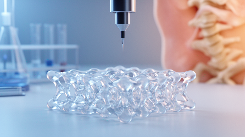
Artificial intelligence is quietly reshaping one of regenerative medicine’s most promising frontiers: the design and printing of hydrogel scaffolds that can help damaged skin and bone to heal. A review published on 30 July 2026 in Frontiers in Biomaterials Science brings together the last five years of research at the intersection of artificial intelligence, 3D printing and hydrogel-based tissue engineering, with a particular focus on musculoskeletal and skin conditions. The article, authored by Kristina Hetherington, Yashaswini Premjit, Nanda Rohra, Merin Lawrence and Payal Ganguly, argues that combining these technologies could move the field away from one-size-fits-all surgical endpoints and towards minimally invasive, patient-specific regenerative therapies.
The review’s starting point is a familiar clinical problem. Conditions that require tissue regeneration, from dermal injuries to musculoskeletal diseases, represent significant global healthcare burdens, and their prevalence rises as populations age. Current clinical approaches to repairing or replacing damaged tissues remain limited by donor shortages, immune rejection and poor long-term integration, and traditional approaches often fail to replicate the complex physiological microenvironments required for functional regeneration. Tissue engineering and regenerative medicine combine biomaterials, cells and molecular signals to create constructs or scaffolds that restore, maintain and enhance tissue function, and hydrogels have emerged as a particularly promising tool for skin and bone applications over the last few decades.
Three-dimensional printing and bioprinting have transformed this landscape by enabling precise, customisable fabrication of scaffolds and hydrogels. Unlike traditional scaffold fabrication methods, 3D bioprinting allows optimised cellular arrangements, biomaterials and growth factors, facilitating the creation of biomimetics that better mimic the physiology of the target tissue both structurally and functionally. The method has demonstrated reproducibility, adaptability towards patient-specific design, and the possibility of integrating multiple cell types within heterogeneous microenvironments. Artificial intelligence now accelerates this progress by addressing design optimisation, process control and quality assurance: AI-driven algorithms can enable predictive modelling of mechanical performance and cell viability, reducing trial-and-error experimentation and optimising bioink formulations, while machine learning approaches have been applied to automate print parameter selection, improve structural accuracy and detect defects in real time.
For bone, the review situates this technology against the biology of a remarkably dynamic organ. Bone healing progresses through inflammation, soft callus formation, hard callus formation and remodelling, with bone marrow mesenchymal stem and stromal cells differentiating into chondrocytes and osteoblasts to form new matrix before mineralisation restores strength and function. When healing is impaired by degenerative disease or trauma, large defects, non-unions and complications follow, and traditional autografts and allografts face donor-site morbidity, limited graft availability, immune rejection and inconsistent outcomes. The authors describe the ‘diamond concept’ framework for bone tissue engineering, which combines a mechanical substrate, osteoconductive scaffolds, osteogenic cells such as mesenchymal stromal cells, and growth factors including bone morphogenic protein 2 and vascular endothelial growth factor to enable bone formation.
Recent studies compiled in the review show how far 3D-printed hydrogel systems for bone have advanced since 2020. Gelatin methacrylate (GelMA) scaffolds modified with luteolin-loaded ZIF-8 nanoparticles demonstrated osteogenic, immunomodulatory and antibacterial activity in a rat model in vivo. Other printed systems include strontium-doped hydroxyapatite in GelMA printed with human fetal osteoblasts, cerium-doped bioactive glass composites tested in rat cranial defects, and scaffolds delivering sequential VEGF and BMP-2 release that promoted early vascularisation followed by osteogenesis in critical-size rat cranial defects. A GelMA composite hydrogel with an iron-chelating network activated HIF-1α to promote chondrogenesis, angiogenesis and vascularised bone regeneration through endochondral ossification. The authors note that while the exact mechanistic pathways of these studies are yet to be explored, the evidence demonstrates strong potential for enhanced bone repair and regeneration.
Artificial intelligence is already contributing to bone tissue engineering in several concrete ways, though the review is careful to distinguish what has been demonstrated from what remains aspirational. Nadernezhad and colleagues employed an interpretable machine learning framework using rheological descriptors from 180 hydrogel formulations to determine key characteristics associated with printability, showing that printability is best understood through collaborative rheological assessments rather than a single universal threshold. Chen and colleagues built decision-tree, random-forest and deep-learning models from 210 direct-ink-writing formulations containing 16 biomaterials and four solvents, with all models achieving accuracies exceeding 80 percent and the random forest performing best. Mohammad and colleagues trained a multilayer perceptron to predict the storage and loss moduli of 3D-printed polyacrylamide hydrogels from seven formulation variables, then used variational autoencoder models to generate candidate material compositions for target rheological outputs.
Beyond formulation screening, AI has been applied to the printing process itself and to scaffold architecture. Bozorg and colleagues combined design of experiments, image-based characterisation and machine learning to study pneumatic semisolid extrusion of model biomedical gels, finding that machine learning may offer a more flexible framework than conventional design of experiments alone for identifying robust operating windows. Rafieyan and colleagues presented an open-source dataset of 1,171 3D-printed scaffolds with machine learning techniques to predict printability, scaffold integrity and cellular response. For architecture, Liu and colleagues introduced a back-propagation neural network with a regenerative genetic algorithm for the inverse design of anisotropic TPMS bone scaffolds, reporting average errors of less than 3 percent for three design targets and approximately 5 percent for six, while Wang and colleagues demonstrated a data-driven inverse-design framework generating spinodoid scaffolds whose elastic constants closely aligned with target bone. Li and colleagues recently created a deep-learning model, OIODNet, that predicts the osteogenic potential of orthopaedic implant surfaces based on early cell morphology. Yet the review stresses that comparatively few studies have employed AI to predict bone regeneration outcomes such as osteogenesis, vascularisation or defect healing in vivo, and that current primary outcomes remain mechanical, structural or mechanobiological rather than definitive evidence of improved bone healing.
For skin, the challenges are of a different character. The review describes the skin as the largest organ of the human body, complex and anisotropic, whose function depends on the highly ordered arrangement of secondary protein structures within its extracellular matrix. Collagen accounts for 70 to 80 percent of the skin’s dry weight, with Type I and Type III collagen giving skin its mechanical resilience along natural tension lines known as Langer’s Lines, and advances in AI-driven path planning now allow hydrogel microstructures to be aligned with these lines. The epidermis is an avascular layer dominated by keratinocytes, the dermis is populated by fibroblasts embedded in dense extracellular matrix, and the hypodermis serves as a reservoir for adipose-derived stem cells that promote growth factors such as VEGF via paracrine secretion. Traditional skin grafts, the review notes, act as patches rather than true replacements, causing donor-site morbidity and failing to regenerate appendages like sweat glands and hair follicles, while chronic wounds such as diabetic foot ulcers cost the US healthcare system over $25 billion annually as of 2026. A 2025 study achieved a three-dimensional gelatin methacryloyl hydrogel containing stratified epidermal cells, fibroblasts and human umbilical vein endothelial cells with interlayer bonding that closely mimicked native skin structure, though it lacked full protein-protein interaction signalling functionality.
Machine learning is reshaping skin bioprinting in parallel ways. ML models have been used to predict print fidelity and structural outcomes based on bioink composition and processing conditions, reducing experimental iterations, and ML-enabled predictive frameworks applied to microneedle fabrication have demonstrated strong correlations between process parameters and structural features. Closed-loop AI systems integrated with real-time imaging and sensor feedback enable adaptive control of the printing process, minimising defects and enhancing reproducibility, while computer vision and deep learning techniques enable automated assessment of print fidelity, cell distribution and structural integrity. Recent composite biomaterials include GelMA-PEG and collagen-PCL systems with improved printability and stability, decellularised extracellular matrix bioinks that retain native biochemical cues, and nanocomposite hydrogels incorporating silver, gold or graphene oxide nanoparticles that impart antimicrobial properties and enhanced electrical conductivity.
The review closes with a sober assessment of what stands between these technologies and the clinic. Personalisation and scale-up are hindered by complex design spaces and poor reproducibility, technical limitations in sensing and automation reduce fidelity, and vascularisation, immune compatibility and regulatory challenges delay clinical translation. Encapsulated cells and bioactive molecules often exhibit reduced stability and inconsistent release profiles within hydrogel matrices, and fermentation variability for polysaccharide-derived bioinks introduces batch-to-batch inconsistencies in molecular weight, purity and rheological behaviour. The authors call for standardised datasets and multi-modal data integration of bone and skin microenvironments, AI-driven Quality by Design frameworks and digital twins, and anticipate autonomous bioprinting with real-time monitoring. Their overall message is that AI in bone-related 3D printing is most established in formulation screening, process optimisation and scaffold design, while its application to direct prediction of regenerative outcomes remains at an earlier stage, making interdisciplinary collaboration the decisive next step towards data-driven, patient-specific regenerative therapies.
Subject of Research: The intersection of artificial intelligence and 3D-printed hydrogels in tissue engineering and regenerative medicine for skin and bone.
Article Title: Hydrogels and artificial intelligence (AI) in tissue engineering and regenerative medicine (TERM): an update on ‘skin and bones’
Article References: Hetherington, K., Premjit, Y., Rohra, N., Lawrence, M., & Ganguly, P. (2026). Hydrogels and artificial intelligence (AI) in tissue engineering and regenerative medicine (TERM): an update on ‘skin and bones’. Frontiers in Biomaterials Science, 5, Article 1886858. https://doi.org/10.3389/fbiom.2026.1886858
Image Credits: AI Generated
DOI: 10.3389/fbiom.2026.1886858
Keywords: tissue engineering, regenerative medicine, hydrogels, 3D bioprinting, artificial intelligence, machine learning, bone regeneration, skin regeneration, bioinks, scaffolds, precision medicine, biomaterials
Cite Scienmag News
Denise Maddox. (October 27, 2026). AI-Designed 3D-Printed Hydrogels Move Toward Repairing Skin and Bone. Scienmag. https://scienmag.com/ai-designed-3d-printed-hydrogels-move-toward-repairing-skin-and-bone/
Denise Maddox. "AI-Designed 3D-Printed Hydrogels Move Toward Repairing Skin and Bone." Scienmag, 27 October 2026, https://scienmag.com/ai-designed-3d-printed-hydrogels-move-toward-repairing-skin-and-bone/. Accessed 27 October 2026.
Denise Maddox. "AI-Designed 3D-Printed Hydrogels Move Toward Repairing Skin and Bone." Scienmag. October 27, 2026. https://scienmag.com/ai-designed-3d-printed-hydrogels-move-toward-repairing-skin-and-bone/


