


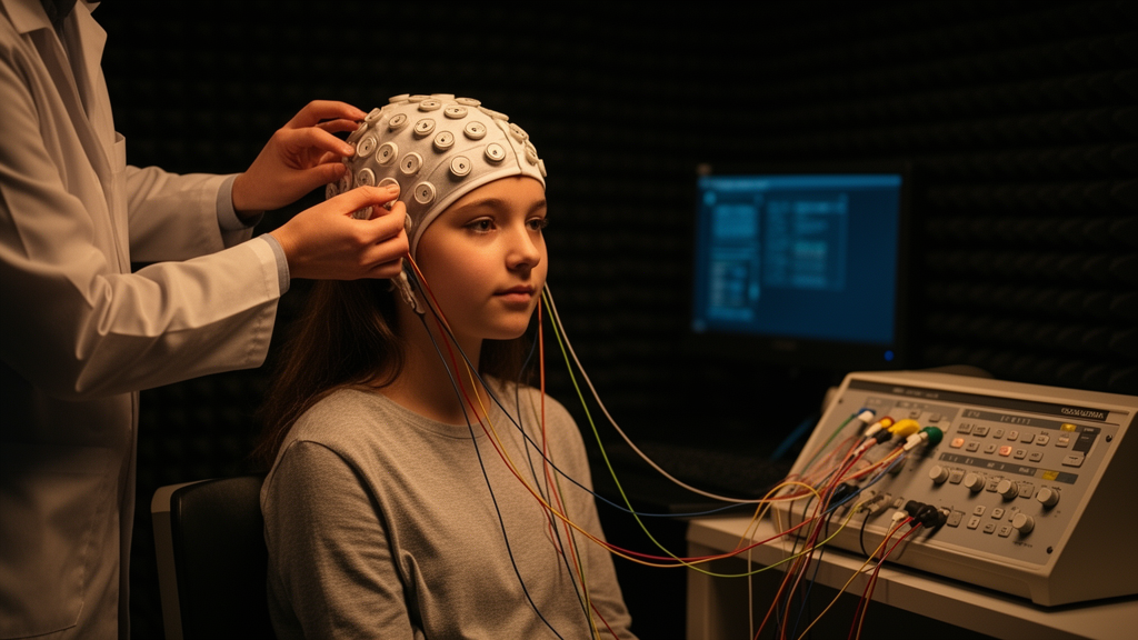
Adolescents with elevated depressive symptoms show a distinctive pattern of brain network alterations that only becomes fully visible when researchers analyze resting-state EEG across multiple spatial scales, according to a new study published in Frontiers in Psychiatry. The research, led by Linfeng Hou and colleagues at Shaanxi Normal University in Xi’an, China, recorded six minutes of eyes-closed resting-state EEG from 96 adolescents with depressive symptoms and 154 healthy controls, then applied an unusually comprehensive analytical framework spanning whole-brain, regional, and single-electrode levels. The findings suggest that early depressive symptoms are associated not with simple global network breakdown, but with a coexisting pattern of compensatory hyperconnectivity, reduced dynamic flexibility, and localized topological vulnerability that coarser analyses would miss.
The study addressed two persistent methodological problems in EEG connectomics. First, traditional network construction relies on fixed thresholds—either absolute cutoffs or proportional rules that force every participant’s network to have the same connection density. Proportional thresholds can mask genuine between-group differences in connectivity density, while fixed absolute thresholds can fragment the networks of participants with weak baseline connectivity into non-physiological shapes. To avoid these distortions, the team used a data-driven adaptive thresholding strategy that determined individualized thresholds from the distribution of each participant’s phase lag index (PLI) connectivity matrix. The PLI metric itself was chosen because it ignores connections with zero phase difference, making it robust against false connectivity estimates caused by volume conduction and common referencing. The adaptive approach identified the most significant connections using eigenvector centrality and calibrated each participant’s threshold against a healthy reference baseline.
The researchers validated their constructed networks using dual small-world normalization metrics, SW1 and SW2, which compare each network against both random and lattice null models. Across all four frequency bands—delta (1–4 Hz), theta (4–7 Hz), alpha (8–13 Hz), and beta (14–30 Hz)—the networks of both groups robustly satisfied small-world criteria, with SW1 medians clustering near 0.94 to 0.95. Notably, the adaptive threshold itself was significantly higher in the depressive-symptoms group only in the theta band, while network density showed no significant between-group differences in any band, ensuring that subsequent topological comparisons were made over comparable network backbones rather than artifacts of unequal sparsity.
At the macroscopic, whole-brain level, the most striking finding was theta-band hyperconnectivity. The adolescents with depressive symptoms showed significantly elevated global static functional connectivity in the theta band compared with controls (controls: 0.181 ± 0.007; symptom group: 0.189 ± 0.017; corrected p < 0.001), while delta and alpha connectivity remained unchanged. A beta-band difference observed before correction did not survive multiple-comparison correction. The team then applied dynamic functional connectivity analysis, using principal component reduction and k-means clustering to identify three recurring whole-brain connectivity states. Across all frequency bands, the symptom group made significantly fewer state transitions than controls, indicating reduced dynamic flexibility. Theta-band dwell time in the baseline-like state was shortened, beta-band dwell time in a third state was prolonged, and beta-band Markov transition probabilities showed increased self-persistence of states alongside reduced return to the baseline state.
Notably, higher-order topological measures at the whole-brain level showed no significant between-group differences. Persistent entropy and landscape-function norms derived from topological data analysis failed to distinguish the groups after correction for multiple comparisons. The authors interpret this as a spatial masking effect: if early network abnormalities are confined to specific nodes, edges, or local subnetworks, whole-brain averages may dilute these focal anomalies alongside compensatory changes elsewhere. This masking effect motivated the descent to finer scales. All initially significant macroscopic findings survived a stringent 100,000-iteration permutation test and bootstrap validation, with confidence intervals excluding zero.
At the mesoscopic level, the theta-band hyperconnectivity was localized to a network centered on the central region, which roughly corresponds to sensorimotor cortex. Connectivity within the central region and between the central region and frontal, parietal, and temporal lobes was significantly elevated in the symptom group, with effect sizes ranging from 0.235 to 0.314. In the alpha band, the symptom group showed focal hypoconnectivity within the occipital region and between occipital and parietal regions, a finding the authors interpret cautiously as possibly reflecting altered sensory gating during relaxed wakefulness. Dynamic connectivity showed a different pattern: the only significant mesoscopic finding was increased temporal variability of frontal-frontal connections in the delta band, which survived supplementary validation using absolute standard deviation, indicating the effect was not an artifact of baseline connectivity differences. Region-averaged topological measures again showed no significant differences, reinforcing the need for finer spatial resolution.
The microscopic, electrode-level analysis revealed the most spatially specific abnormalities. After correction across 1,740 edge tests, static theta-band hyperconnectivity was confined to just two connections along the midline prefrontal-to-central axis: the left frontal pole to midline central region (Fp1–FCz) and the midline prefrontal to midline central connection (Fz–FCz). Dynamic connectivity abnormalities, corrected across the same edge space, were confined to theta-band pathways running from the right prefrontal cortex to the midline occipital cortex, specifically Fp2–Oz and F4–Oz, indicating instability in how right prefrontal regulatory systems fluctuate in their coupling with visual regions during rest. Node-level topological analysis, based on persistent homology, showed reduced node frequency and node persistence at the right prefrontal electrodes Fp2 and F4 in the theta band, suggesting weakened local topological support for higher-order information integration at these hubs. The left temporal node FT7 showed increased topological metrics in the alpha band, an isolated exploratory finding the authors link cautiously to language and semantic-emotional processing.
To integrate these findings, the researchers constructed a multidimensional convergence matrix mapping the spatial overlap between network abnormalities and the approximate scalp projections of prefrontal neuromodulation sites used in non-invasive brain stimulation research. All five prefrontal electrodes examined—Fp1, Fp2, F3, Fz, and F4—showed theta-band abnormalities, but with distinct feature profiles. Fp1, F3, and Fz showed abnormalities only in static connectivity, while Fp2 and F4 showed convergent abnormalities in the two related topological indices, which the authors note were strongly correlated at these nodes and should be interpreted as one related feature profile rather than two independent findings. Beyond the traditional prefrontal sites, the analysis flagged three additional nodes warranting further investigation: FCz, the only static-connectivity node reaching high significance, potentially anchoring a sensorimotor-somatization axis; Oz, the only node showing dynamic-connectivity abnormality, possibly related to visual information regulation; and FT7, with its exploratory alpha-band topological profile.
The authors emphasize that these mappings are exploratory computational clues rather than validated stimulation targets. The 10–20 EEG electrode positions represent approximate scalp-surface projections, not precise cortical targets, and clinical transcranial magnetic stimulation relies on MRI-guided neuronavigation to account for individual neuroanatomy. No intervention was performed in this study, so the spatial overlap cannot be interpreted as evidence of therapeutic efficacy. The authors also caution that frontopolar and midline electrodes are susceptible to residual ocular and myogenic artifacts, and that the two-second analysis windows contain relatively few oscillatory cycles in the delta band, warranting cautious interpretation of low-frequency dynamic findings—though supplementary validations supported the delta-band frontal result.
Several further limitations frame the conclusions. The cross-sectional design precludes causal conclusions about the sequence of network changes. The symptom group was defined by threshold scores on two self-report scales, the CES-D and PHQ-9, combined with review by school counseling professionals, rather than structured diagnostic interviews, so the findings represent EEG correlates of elevated depressive symptoms rather than biomarkers of clinically diagnosed major depressive disorder. No psychiatric clinical control groups were included, leaving open whether the observed alterations are specific to depressive symptoms or reflect broader transdiagnostic neural correlates. The authors frame their compensatory interpretation of theta hyperconnectivity as hypothesis-generating, and call for future studies incorporating structured diagnostics, longitudinal designs, high-density EEG source localization, EEG-fMRI multimodal data, and intervention trials to validate the neural origins and clinical translational value of these multiscale network features.
Subject of Research: Multiscale resting-state EEG network alterations in adolescents with depressive symptoms
Article Title: Multiscale EEG network alterations in adolescents with depressive symptoms: static–dynamic connectivity, topological features, and exploratory scalp-level network hubs
Article References: Hou, L., Chen, M., Yang, J., Ren, Y., Peng, T., & Li, Y. (2026). Multiscale EEG network alterations in adolescents with depressive symptoms: static–dynamic connectivity, topological features, and exploratory scalp-level network hubs. Frontiers in Psychiatry, 17, Article 1943613. https://doi.org/10.3389/fpsyt.2026.1943613
Image Credits: AI Generated
DOI: 10.3389/fpsyt.2026.1943613
Keywords: EEG, adolescent depression, brain networks, functional connectivity, theta band, topological data analysis, dynamic functional connectivity, adaptive thresholding, phase lag index, neuromodulation, prefrontal cortex, resting state
Cite Scienmag News
Cassandra Pierce. (October 27, 2026). EEG Reveals Hidden Brain Network Signatures in Adolescents with Depressive Symptoms. Scienmag. https://scienmag.com/eeg-reveals-hidden-brain-network-signatures-in-adolescents-with-depressive-symptoms/
Cassandra Pierce. "EEG Reveals Hidden Brain Network Signatures in Adolescents with Depressive Symptoms." Scienmag, 27 October 2026, https://scienmag.com/eeg-reveals-hidden-brain-network-signatures-in-adolescents-with-depressive-symptoms/. Accessed 27 October 2026.
Cassandra Pierce. "EEG Reveals Hidden Brain Network Signatures in Adolescents with Depressive Symptoms." Scienmag. October 27, 2026. https://scienmag.com/eeg-reveals-hidden-brain-network-signatures-in-adolescents-with-depressive-symptoms/


