


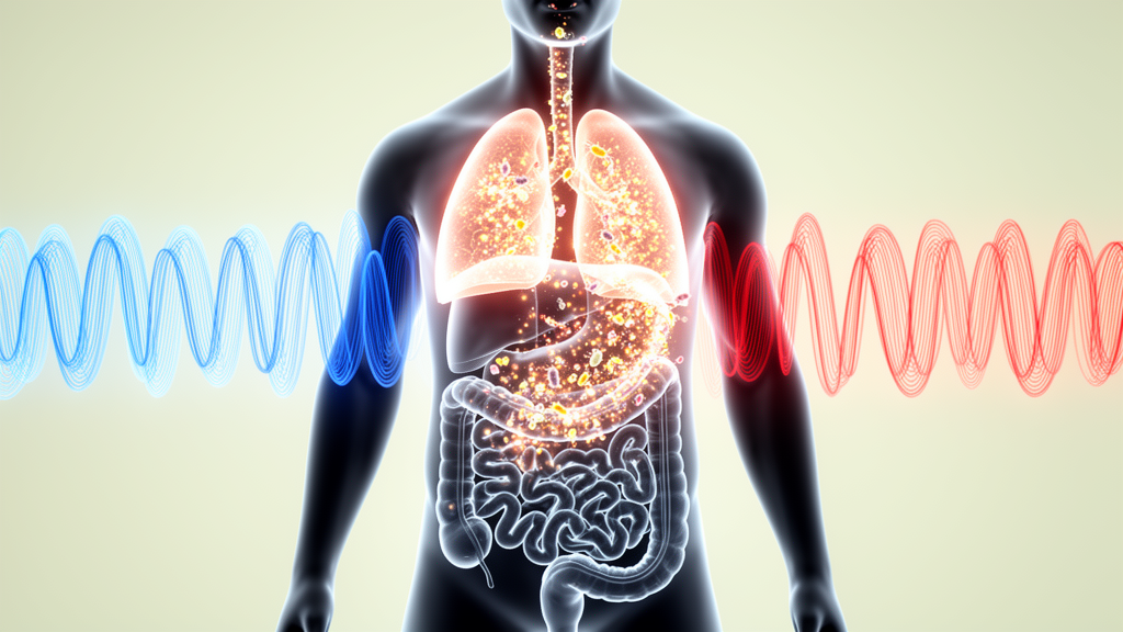
Obstructive sleep apnea affects an estimated 936 million adults aged 30 to 69 worldwide, including roughly 425 million with moderate-to-severe disease, and its consequences extend far beyond a night of disrupted breathing. Each apneic episode subjects the body to intermittent hypoxia, a cyclical pattern of oxygen deprivation and reoxygenation that fuels oxidative stress, systemic inflammation, endothelial dysfunction, and metabolic dysregulation. A new narrative review published in Frontiers in Cellular and Infection Microbiology argues that one of the most overlooked battlegrounds in this process is the gut, where recurrent oxygen swings remodel the microbial ecosystem, weaken the intestinal barrier, and alter a network of host-microbial metabolites that may help drive injury in the heart, liver, brain, and kidneys.
The review, led by researchers at Lishui Hospital of Traditional Chinese Medicine affiliated with Zhejiang University of Chinese Medical University, systematically distinguishes preclinical experimental findings from human clinical observations, an approach that matters because the two bodies of evidence point in different directions. The authors frame intermittent hypoxia as a quantifiable ecological selection pressure acting on the intestinal ecosystem, and they grade the evidence linking microbial changes to organ injury across a spectrum from simple association to phenotype transfer, microbiota dependence, and pathway-specific mechanistic validation. Their central conclusion is deliberately cautious: the gut microbiota is best regarded as a context-dependent contributor and amplifier of hypoxia-related injury, not a universal mediator.
The biological logic begins with the colon’s unusual oxygen landscape. In a healthy gut, a radial oxygen gradient falls from the well-perfused mucosal tissue toward a nearly anaerobic luminal core, a structure maintained largely by colonocytes that consume enormous amounts of oxygen as they burn microbiota-derived butyrate through mitochondrial beta-oxidation and oxidative phosphorylation. This physiological hypoxia stabilizes hypoxia-inducible factor, a transcriptional regulator that supports tight-junction integrity, mucus production, and tissue repair. When butyrate production or epithelial oxygen consumption falters, oxygen availability rises at the mucosal-luminal interface, favoring facultative anaerobes, while inflammation-derived nitrate can further fuel the growth of Enterobacteriaceae. The intestinal oxygen environment is thus a dynamic property jointly maintained by mucosal perfusion, epithelial metabolism, and microbial fermentation, and it is precisely this balance that intermittent hypoxia disrupts.
Experimental work shows that the cyclical oxygen fluctuations of sleep apnea can be transmitted to the intestinal wall. In a key study, oxygen tension in fecal material close to the mucosa fluctuated in parallel with changes in inspired oxygen concentration, and prolonged intermittent hypoxia exposure was accompanied by marked alterations in microbial diversity and community structure. Multi-omics studies using models that combine intermittent hypoxia with hypercapnia, the carbon dioxide retention that accompanies real apneas, revealed that microbial remodeling occurs alongside coordinated changes in bile acids, fatty acids, aromatic compounds, and lipid metabolites, with some features reproduced across different rodent models. Notably, some microbial abnormalities and elevated circulating endotoxin levels persist even after normal oxygenation is restored, suggesting that ecological recovery lags behind the normalization of blood gases.
Intermittent hypoxia also damages the gut directly. Repeated hypoxia-reoxygenation cycles enhance oxidative stress, induce mitochondrial injury in intestinal epithelial cells, and activate inflammatory signaling through pathways such as NF-kappaB. Animal studies have documented villous and microvillous injury, reduced goblet cell numbers, disrupted tight-junction architecture, and increased intestinal permeability. Once the barrier is compromised, microbial components such as lipopolysaccharide can enter the portal and systemic circulation and amplify inflammation through TLR4-related pathways, while the resulting inflammatory nitrate promotes the very facultative anaerobes that thrive in a destabilized gut, creating a self-reinforcing loop among dysbiosis, barrier failure, and inflammation. Clinical studies have reported elevated levels of intestinal fatty acid-binding protein, D-lactate, and lipopolysaccharide-binding protein in subsets of patients with sleep apnea, correlating with disease severity and nocturnal hypoxemia, though these markers are also influenced by obesity, diet, and medication, and most studies are cross-sectional.
The strongest causal evidence involves the cardiovascular system, and it comes almost entirely from animal models. In a mechanically induced sleep apnea model, exposure alone under a standard diet did not produce sustained hypertension, but the combination of apnea-like exposure and a high-fat diet caused gut dysbiosis, depletion of short-chain fatty acid-producing bacteria, and elevated blood pressure. Broad-spectrum antibiotics prevented hypertension from developing, and transferring cecal microbiota from apnea-exposed animals to recipients that remained exposed reproduced the hypertensive response. Supplementation with Clostridium butyricum, resistant starch, or directly with the short-chain fatty acid acetate prevented blood-pressure elevation and intestinal inflammation in high-fat-diet-fed rats. Separately, microbiota from donors exposed to isolated intermittent hypoxia transferred elevated blood pressure and impaired aortic and coronary vasorelaxation to normoxic recipients, accompanied by reduced acetate and increased trimethylamine N-oxide, a microbiota-dependent metabolite linked to enhanced platelet reactivity and thrombosis potential.
Immune mechanisms add another layer of specificity. In mechanical apnea models, reduced intestinal regulatory T cells, enhanced Th17 responses, and migration of gut-derived helper T cells have been observed, and neutralization of the inflammatory cytokine IL-17A attenuated blood-pressure elevation and inflammation. In the neurocognitive domain, a model combining intermittent hypoxia with a ketogenic diet showed that the exposure enriched Bilophila wadsworthia and aggravated learning and memory impairment; microbiota depletion attenuated the injury, while transferring the altered microbiota or colonizing animals with B. wadsworthia alone induced impaired hippocampal long-term potentiation, reduced neurogenesis, and cognitive decline, with inhibition of Th1-cell development preventing these deficits. In a complementary finding, loss of Bifidobacterium pseudolongum during chronic intermittent hypoxia was linked to cognitive impairment, and supplementation with the bacterium or with acetate restored circulating acetate, reduced microglial activation, suppressed cGAS-STING signaling and neuronal PANoptosis in the hippocampus, and improved performance.
Metabolic and hepatic evidence follows a similar but less complete trajectory. Microbiota transfer from hypoxia-exposed donors induced barrier abnormalities and insulin resistance in normoxic recipients, and plasma extracellular vesicles from hypoxia-exposed animals, and from patients with sleep-disordered breathing, suppressed insulin-stimulated AKT phosphorylation in adipocytes, though the intestinal origin of these vesicles remains unestablished. In the liver, eight weeks of chronic intermittent hypoxia in lean mice on standard chow predominantly induced metabolic and transcriptional changes, while sixteen weeks produced glucose abnormalities, lipid accumulation, and inflammation; broad-spectrum antibiotics partially attenuated the steatosis, and the exposure was associated with depletion of Bacteroides uniformis and elevation of the bile acid deoxycholic acid alongside altered PPAR-related signaling. The authors caution, however, that the study did not demonstrate that loss of B. uniformis caused the bile acid changes, that antibiotics have microbiota-independent effects, and that germ-free attenuation of atherosclerosis in a related model was confounded by both high-fat diet and hypercapnia.
By contrast, the evidence for kidneys, heart muscle, and other organs remains largely associative. No study has yet shown that microbiota from apnea- or hypoxia-exposed donors can reproduce a renal injury phenotype in unexposed recipients, and gut-kidney mechanisms are inferred mainly from chronic kidney disease literature involving metabolites such as indoxyl sulfate, p-cresyl sulfate, and trimethylamine N-oxide, findings that cannot be directly extrapolated because circulating metabolite concentrations are themselves shaped by renal clearance. For the myocardium, germ-free and conventionally colonized mice show different left ventricular transcriptional responses to combined hypoxia and hypercapnia, but there is no evidence that the microbiota can transfer or reverse structural or functional heart injury. The review’s cross-organ comparison therefore places hypertension at the top of the evidence hierarchy with convergent preclinical functional support, vascular dysfunction and insulin resistance at the functional level, neurocognitive injury at the mechanistically supported but model-limited level, and atherosclerosis, hepatic steatosis, myocardial, renal, and other phenotypes progressively lower.
The clinical implications remain open. Most human studies are cross-sectional, making it impossible to determine whether microbiome abnormalities cause organ injury, result from it, or merely correlate with shared factors such as obesity. Large-scale metagenomic work suggests that hypoxemia-related parameters, including time spent with oxygen saturation below 90 percent and the oxygen desaturation index, associate more strongly with microbial species and functional pathways than the apnea-hypopnea index alone, echoing cohort findings that integrated hypoxic burden predicts cardiovascular events better than event frequency. Continuous positive airway pressure offers a natural test of reversibility, but current findings are inconsistent, with some studies showing little microbial recovery after treatment and metagenomic analyses reporting compositional and pathway changes in severe disease. The authors call for longitudinal cohorts of untreated patients, paired intestinal and circulating metabolite measurements, standardized microbiome protocols, and precision interventions targeting defined strains, metabolites such as acetate, or host receptors including FXR, TGR5, and short-chain fatty acid receptors, always as adjuncts to established therapy rather than substitutes. Until such studies are done, the gut microbiota stands as a promising but unproven amplifier of sleep apnea’s systemic toll, its importance varying with exposure pattern, diet, obesity, and the vulnerability of each target organ.
Subject of Research: Gut microbiota and metabolic remodeling under intermittent hypoxia in obstructive sleep apnea
Article Title: Intermittent hypoxia–driven gut microbial and metabolic remodeling in obstructive sleep apnea: mechanisms and evidence across target organs
Article References: Wu, M., Chen, L., Zhou, J., Huang, H., Chen, X., Wan, S., Xie, X., Zhou, M., & Zhang, Z. (2026). Intermittent hypoxia–driven gut microbial and metabolic remodeling in obstructive sleep apnea: mechanisms and evidence across target organs. Frontiers in Cellular and Infection Microbiology, 16, Article 1965973. https://doi.org/10.3389/fcimb.2026.1965973
Image Credits: AI Generated
DOI: 10.3389/fcimb.2026.1965973
Keywords: obstructive sleep apnea, intermittent hypoxia, gut microbiota, short-chain fatty acids, bile acids, TMAO, intestinal barrier, hypertension, hepatic steatosis, neurocognitive impairment, microbiota transplantation, gut-liver axis
Cite Scienmag News
Morgan Morrow. (October 26, 2026). How Sleep Apnea’s Oxygen Swings Reshape the Gut Microbiome and Drive Organ Injury. Scienmag. https://scienmag.com/how-sleep-apneas-oxygen-swings-reshape-the-gut-microbiome-and-drive-organ-injury/
Morgan Morrow. "How Sleep Apnea’s Oxygen Swings Reshape the Gut Microbiome and Drive Organ Injury." Scienmag, 26 October 2026, https://scienmag.com/how-sleep-apneas-oxygen-swings-reshape-the-gut-microbiome-and-drive-organ-injury/. Accessed 26 October 2026.
Morgan Morrow. "How Sleep Apnea’s Oxygen Swings Reshape the Gut Microbiome and Drive Organ Injury." Scienmag. October 26, 2026. https://scienmag.com/how-sleep-apneas-oxygen-swings-reshape-the-gut-microbiome-and-drive-organ-injury/


