


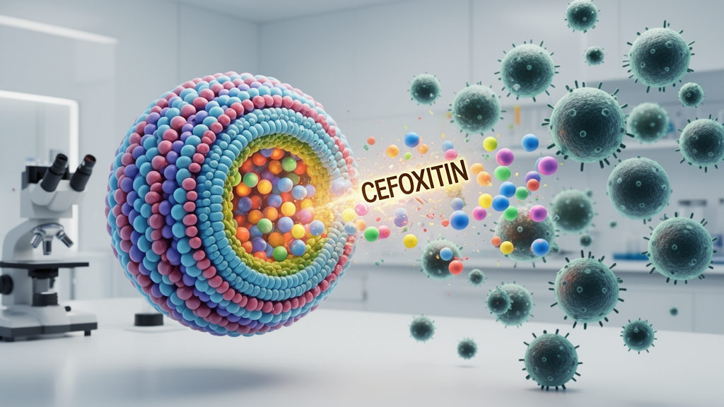
Antibiotic resistance has been described as a silent pandemic of the twenty-first century, and researchers are increasingly turning to drug-delivery engineering rather than new molecules to squeeze more efficacy out of the antibiotics that still work. A new laboratory study published in International Microbiology reports that encapsulating the antibiotic cefoxitin inside niosomes — tiny vesicles built from nonionic surfactants and cholesterol — substantially boosted the drug’s antibacterial and antibiofilm performance against four important bacterial species: Escherichia coli, Acinetobacter baumannii, Staphylococcus aureus, and Listeria monocytogenes. The work, led by Masoud Faraji, Saeedeh Keyvani-Ghamsari, and Amir Mirzaie, is entirely in vitro, but its results offer a concrete example of how nanoscale packaging can change the behavior of an established antibiotic.
Cefoxitin itself is a semi-synthetic, second-generation cephalosporin derived from cephamycin C, delivered by injection to treat serious infections of the urinary tract, bloodstream, bones and joints, the lower respiratory tract, and the gastrointestinal tract. It is also widely used as a prophylactic before surgery to prevent post-surgical infections, and Health Canada classifies it as an antibiotic of “high importance” for managing multiple infectious diseases. The drug has shown low toxicity in humans. Yet cefoxitin-resistant strains of all four bacteria studied here have been repeatedly identified, driven by mechanisms that include increased expression of the chromosomal ampC gene, acquisition of beta-lactamase genes from other bacteria, efflux pumps that lower intracellular drug concentrations, mutations in the ompK35 and ompK36 porin channels that reduce drug penetration, and the formation of biofilms.
Biofilms are central to the problem the researchers set out to address. These thin layers of bacteria attach to living or non-living surfaces and are wrapped in a polymer matrix of polysaccharides, proteins, and nucleic acids that confers high resistance to both host immune responses and antibiotic treatment. Biofilm-associated infections have been estimated to account for roughly 65 to 80 percent of all microbial infections, and the study’s authors note that infections caused by multidrug-resistant bacteria rank as the third leading cause of death worldwide, after cardiovascular diseases. Compounding the challenge, antibiotics often suffer from low bioavailability and limited penetration into sites of bacterial growth, further undermining treatment outcomes. Niosomes, the authors argue, are one of the most promising nanocarriers for targeted delivery: they are composed of amphipathic lipids and nonionic surfactants in single-layered or multi-layered form, can encapsulate both hydrophilic and lipophilic compounds, and offer biocompatibility, degradability, low toxicity, low production cost, non-immunogenicity, and flexible storage.
The team prepared cefoxitin-loaded niosomes by thin-layer hydration. A lipid phase containing cholesterol, Span 60, and Tween 60 at a molar ratio of 1:1:1 was dissolved in a chloroform–methanol mixture, with the total molar amount of the three components relative to cefoxitin set at 30:1. A rotary evaporator removed the solvent under vacuum at 60 degrees Celsius, leaving a thin lipid film that was then hydrated with phosphate buffer containing cefoxitin at 1 milligram per milliliter. Sonication for seven minutes reduced the particle size. The standard strains used — S. aureus ATCC 33591, E. coli ATCC 25922, A. baumannii ATCC 19606, and L. monocytogenes ATCC 19115 — came from the microbial bank of the Pasteur Institute of Iran.
Characterization showed a highly efficient encapsulation process. The calculated entrapment efficiency of cefoxitin was 89.36 percent, determined by centrifuging the formulation at 14,000 times gravity for 45 minutes at 4 degrees Celsius and measuring the free drug left in the supernatant by UV–Vis spectrophotometry at 235 nanometers. Scanning electron microscopy revealed well-dispersed, predominantly spherical vesicles consistent with successful formation. Dynamic light scattering reported a Z-average particle size of 373.9 nanometers, while the size distribution by number showed a major peak at 142.3 nanometers accounting for 97.7 percent of the measured distribution, with a minor peak at 948 nanometers making up the remaining 2.3 percent. The polydispersity index was 0.526, indicating a relatively broad size distribution and heterogeneity within the vesicle population — a value the authors say suggests further formulation optimization may be required to achieve a more homogeneous system.
Fourier transform infrared spectroscopy provided evidence that the drug survived encapsulation chemically intact. The spectrum of free cefoxitin showed a strong stretching band in the 3400 to 3600 per centimeter region consistent with –OH and –NH groups, a distinct band at 1638.99 per centimeter related to the amide groups and beta-lactam ring, and a small peak at 668 per centimeter consistent with out-of-plane bending of the beta-lactam ring or C–S bonds. The spectrum of the loaded niosomes was broadly similar, with slight decreases in intensity and slight shifts of some bands that the researchers attribute to non-covalent interactions such as hydrogen bonding between the drug and the niosomal components. A new small peak in the 1200 to 1300 per centimeter region likely reflects physical interactions between the drug and the vesicle matrix. Together, the spectra indicate that cefoxitin was physically encapsulated without chemical modification.
Release testing revealed the sustained-release behavior that may underpin the formulation’s enhanced activity. In a dialysis setup held at 37 degrees Celsius in phosphate-buffered saline, approximately 80 percent of free cefoxitin was released within the first 8 hours, whereas only about 30 percent of the niosomal drug was released in the same period. By 24 hours, the free drug had reached roughly 98 percent release while the niosomes had delivered about 50 percent. Over the full 72-hour experiment, cumulative release of free cefoxitin approached 100 percent, compared with 62.69 percent from the niosomal formulation. The authors suggest that the combination of Span 60, cholesterol, and Tween 60 contributes to the rigidity and stability of the vesicular structure, slowing the release rate, and note that previous studies have reported higher stability and entrapment efficiency for niosomes made with these longer-chain surfactants.
The antibacterial results were the study’s most striking finding. In well diffusion assays, free cefoxitin showed negligible inhibitory activity against the Gram-negative strains E. coli and A. baumannii, while cefoxitin-loaded niosomes produced clear inhibition zones around the wells. Microdilution testing confirmed the pattern: free cefoxitin failed to inhibit either Gram-negative strain at any concentration tested, from 3.125 to 100 micrograms per milliliter, whereas the niosomal formulation inhibited growth of both strains at 50 micrograms per milliliter. Against the Gram-positive strains, the minimum inhibitory concentration of free cefoxitin for S. aureus and L. monocytogenes was 50 micrograms per milliliter, but the niosomal formulation halved it to 25 micrograms per milliliter. The minimum bactericidal concentration told a similar story: neither formulation was bactericidal against E. coli or A. baumannii at the concentrations tested, but free cefoxitin killed the Gram-positive strains at 100 micrograms per milliliter while the niosomal version achieved the same at 50 micrograms per milliliter.
Biofilm inhibition, measured by crystal violet staining in microtiter plates at half the minimum inhibitory concentration, followed the same trend. Free cefoxitin had little impact on biofilm formation by the Gram-negative bacteria, whereas the niosomal formulation significantly reduced biofilm formation compared with the untreated control. Against the Gram-positive strains, free cefoxitin did significantly reduce biofilm formation relative to the control, but the reduction was more pronounced with niosomal cefoxitin, which produced lower optical density values at 570 nanometers. A 72-hour time-kill assay on S. aureus and E. coli, chosen to represent one Gram-positive and one Gram-negative strain, supported these findings: bacteria exposed to half the minimum inhibitory concentration of niosomal cefoxitin showed lower optical absorbance at 600 nanometers than bacteria exposed to the free drug at every time point measured, from 0 to 72 hours, with the reduction more pronounced in S. aureus than in E. coli.
The authors are careful about what the results do and do not demonstrate. They suggest the enhanced activity may be associated with the sustained release profile, and note that previous studies have linked improved niosomal performance to interactions between nanovesicles and bacterial cells, changes in membrane permeability, or improved drug delivery — but these mechanisms were not directly investigated in the present study and cannot be confirmed from the current findings. Whether the greater effect against Gram-negative bacteria relates to altered drug delivery or to interactions with the bacterial envelope remains unclear, particularly since the presence of an outer membrane may contribute to the lower susceptibility of Gram-negative organisms to free cefoxitin. The study also has clear limitations: the cytotoxicity and biocompatibility of the cefoxitin-loaded niosomes were not evaluated, so the safety of the formulation cannot be assessed from these data, and further optimization of lipid and surfactant ratios may help lower the polydispersity index. The authors call for appropriate cell-based and in vivo models, pharmacokinetic analyses, and kinetic modeling of the release behavior before any clinical conclusions can be drawn. For now, the findings stand as an in vitro demonstration that niosomal encapsulation can enhance both the antibacterial and antibiofilm activity of cefoxitin — a proof of concept that joins a growing body of work showing niosomal formulations of cephalexin, ciprofloxacin, cefazolin, teicoplanin, and nisin–zinc oxide combinations improving the performance of other antimicrobial agents.
Subject of Research: Niosomal encapsulation of the antibiotic cefoxitin and its enhanced in vitro antibacterial and antibiofilm activity against Gram-positive and Gram-negative bacteria
Article Title: Niosomal delivery of cefoxitin enhances antibacterial and antibiofilm activities against gram-positive and gram-negative bacteria
Article References: Faraji, M., Keyvani-Ghamsari, S., & Mirzaie, A. (2026). Niosomal delivery of cefoxitin enhances antibacterial and antibiofilm activities against gram-positive and gram-negative bacteria. International Microbiology. https://doi.org/10.1007/s10123-026-00912-2
Image Credits: AI Generated
DOI: 10.1007/s10123-026-00912-2
Keywords: cefoxitin, niosomes, antibiotic resistance, biofilm, drug delivery, nanovesicles, Escherichia coli, Staphylococcus aureus, Acinetobacter baumannii, Listeria monocytogenes, sustained release, antimicrobial nanotechnology
Cite Scienmag News
Drew Townsend. (October 24, 2026). Niosome-Encapsulated Cefoxitin Shows Stronger Antibacterial and Antibiofilm Activity in Vitro. Scienmag. https://scienmag.com/niosome-encapsulated-cefoxitin-shows-stronger-antibacterial-and-antibiofilm-activity-in-vitro/
Drew Townsend. "Niosome-Encapsulated Cefoxitin Shows Stronger Antibacterial and Antibiofilm Activity in Vitro." Scienmag, 24 October 2026, https://scienmag.com/niosome-encapsulated-cefoxitin-shows-stronger-antibacterial-and-antibiofilm-activity-in-vitro/. Accessed 24 October 2026.
Drew Townsend. "Niosome-Encapsulated Cefoxitin Shows Stronger Antibacterial and Antibiofilm Activity in Vitro." Scienmag. October 24, 2026. https://scienmag.com/niosome-encapsulated-cefoxitin-shows-stronger-antibacterial-and-antibiofilm-activity-in-vitro/


