


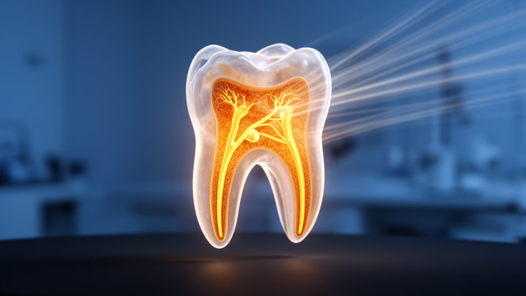
A high-resolution three-dimensional imaging study of children’s extracted baby teeth has revealed just how easy it is for dentists to miss a hidden canal inside the upper primary molars they treat. Using micro-computed tomography, or micro-CT, researchers scanned 60 maxillary primary molars at an isotropic voxel size of 13.61 micrometres, fine enough to resolve structures far smaller than anything visible on a conventional dental radiograph. Their analysis, published as an open-access article in Scientific Reports, found that a second mesiobuccal canal, known as MB2, was present in 43.3 percent of the mesiobuccal roots examined. Whenever that canal existed, its orifice sat palatal to the main mesiobuccal canal, and in most cases it originated in the coronal third of the root. The finding matters because pulpectomy, the common procedure that removes infected pulp tissue from baby molars to keep them functioning until the permanent teeth erupt, depends on locating and cleaning every canal space.
The teeth in the study were collected after routine extractions performed for reasons unrelated to the research, and each had no more than two millimetres of physiological root resorption. The sample comprised 41 primary maxillary first molars and 19 second molars, drawn from children aged roughly four to seven years. Because the teeth were fully anonymised and extracted as part of standard clinical care, the ethics committee waived individual informed consent. Every specimen received a random alphanumeric code so that the single board-certified oral and maxillofacial radiologist who made all measurements remained blinded to specimen identity. Before data collection, the examiner completed a calibration session on independent pilot teeth, and re-analysis of 20 percent of the specimens after a two-week interval showed excellent agreement, with an intraclass correlation coefficient of 0.95.
The scanning protocol itself was unusually detailed. Each tooth was mounted in a custom holder and imaged in a desktop SkyScan 1172 system operating at 85 kilovolts and 118 microamperes, with a rotation step of 0.60 degrees and an exposure time of 2600 milliseconds. Raw projections were reconstructed with beam-hardening correction and ring-artefact reduction, and all quantitative measurements were performed in dedicated analysis software. The researchers standardised their reference levels carefully: cross-sections were taken one millimetre below the canal orifice, at mid-root, and one millimetre coronal to the apical constriction. This non-destructive approach allowed the team to map canal lengths, diameters, inter-orifice distances, dentin wall thickness, isthmuses and accessory canals without cutting into or otherwise altering the precious specimens.
A conservative definition governed what counted as MB2. The researchers required a separate orifice divided from the main mesiobuccal orifice by a complete band of dentin, with the canal space traceable as a continuous lumen for at least two millimetres apical to the orifice. Incompletely separated or shorter canal spaces were classified instead as lateral ramifications. This stricter criterion probably explains why the observed prevalence of 43.3 percent, with a 95 percent confidence interval of 31.6 to 55.9 percent, sits in the middle of the previously reported range of roughly 20 to 60 percent for primary maxillary molars. Of the 26 MB2-positive teeth, the orifice arose in the coronal third in 20 cases and in the middle third in six; none appeared in the apical third.
Three-rooted anatomy dominated the sample, with 50 of the 60 teeth, or 83.3 percent, showing the classic trifurcation into mesiobuccal, distobuccal and palatal roots. The remaining ten teeth, 16.7 percent, displayed the typical two-rooted variant produced by fusion of the distobuccal and palatal roots. Even within those fused roots, both canals remained individually traceable on the micro-CT scans. The prevalence of MB2 in these baby molars is lower than the roughly 60 to 70 percent reported in cone-beam CT studies of permanent maxillary first molars, but the clinical stakes are different: primary molars must survive only a few years, yet untreated canal spaces can harbour microbial biofilms that drive chronic apical periodontitis and treatment failure.
Perhaps the most striking morphometric result concerned canal shape. Across 315 classifiable canal lumens, long-oval cross-sections accounted for 48.3 percent and oval profiles for 45.4 percent, with flattened canals making up just 6.3 percent. Not a single canal met the strict round criterion of an aspect ratio below 1.05. Shape did not differ significantly across the coronal, middle and apical root levels, although oval profiles became proportionally more frequent toward the apex. This matters clinically because rotary nickel-titanium instruments are round in cross-section and therefore cannot fully shape an oval canal, leaving buccal and lingual extensions uninstrumented where biofilm may persist. Conversely, larger or oval-path files risk stripping away dentin in roots whose walls are already dangerously thin.
That danger was quantified directly. The minimum dentin thickness at the apical level was lowest in the palatal roots, averaging just 0.10 millimetres, followed by the mesiobuccal roots at 0.12 millimetres and the distobuccal roots at 0.21 millimetres, a difference that was statistically significant. In other words, the thinnest apical walls in these teeth approach a tenth of a millimetre, leaving very little margin for error during apical preparation. The buccolingual diameter significantly exceeded the mesiodistal diameter in the mesiobuccal, MB2 and distobuccal roots at all three levels, while the palatal root showed no significant directional difference, indicating a more circular cross-section that round instruments can shape more completely.
The inter-orifice measurements added another layer of clinical relevance. The shortest mean separation between any pair of canal orifices was between MB1 and MB2, at just 1.12 millimetres, while the greatest was between MB1 and the palatal canal, at 5.54 millimetres. Such tight proximity substantially increases the risk of overlooking MB2 during access cavity preparation, particularly given its 43.3 percent prevalence. The team also mapped isthmuses, the narrow pulp-filled connections between adjacent canals: these were present in the coronal third in 28.3 percent of specimens, in the middle third in 23.3 percent, and in the apical third in only 6.7 percent, with the distobuccal-palatal region the most frequent site. Accessory canals appeared in all four canal types, most often in the distobuccal root at 43.3 percent and least often in MB2 at 10.0 percent, and were most commonly located in the apical third.
The authors are careful to frame the practical implications as anatomically informed considerations rather than validated clinical recommendations, since no treatment outcomes were tested in this laboratory study. They suggest that magnification with dental loupes or an operating microscope may aid MB2 detection, that circumferential instrumentation may suit the oval mesiobuccal and distobuccal canals, and that apical preparation should be conservative in the palatal and mesiobuccal roots where the walls are thinnest. Cone-beam CT is not a realistic clinical option for primary molars because of radiation considerations and the teeth’s limited remaining lifespan, so the value of this ex vivo characterisation lies in supplying the prior anatomical knowledge clinicians need during conventional treatment.
The study has clear limitations. Its in vitro design cannot be extrapolated directly to clinical outcomes, rare configurations such as four-rooted molars may be under-represented in a sample of 60 teeth, and micro-CT findings do not transfer directly to the two-dimensional radiographs used in everyday practice. The specimens came from two universities in Egypt and do not represent the entire Egyptian population, and because the sample contained markedly unequal numbers of first and second molars, a robust tooth-type comparison was not feasible. The authors call for future work correlating these three-dimensional anatomical findings with clinical pulpectomy outcomes and validating the MB2 diagnostic criteria across multiple operators and imaging systems. Even so, the message for paediatric dentistry is vivid: inside every small baby molar lies a landscape of hidden canals, ribbon-like isthmuses and paper-thin walls that only high-resolution imaging could reveal.
Subject of Research: Three-dimensional root and canal anatomy of maxillary primary molars assessed by micro-computed tomography
Article Title: Micro-computed tomography analysis of root and canal morphology in maxillary primary molars
Article References: Eldehna, A. M., Elzahar, S., Ahmad, A. H., El Kharadly, D. Y., Salem, N. A., Abdel-Ghany, D. M., Abd-Allah, N. F., Abd-Allah, M. F., Ahmed, H. M. A., & Abdelkafy, H. (2026). Micro-computed tomography analysis of root and canal morphology in maxillary primary molars. Scientific Reports, 16(1), Article 33592. https://doi.org/10.1038/s41598-026-71739-0
Image Credits: AI Generated
DOI: 10.1038/s41598-026-71739-0
Keywords: micro-CT, primary molars, MB2 canal, root canal anatomy, pulpectomy, paediatric dentistry, dentin thickness, isthmus, accessory canals, canal morphology, dental imaging, Micro-computed
Cite Scienmag News
Harold Sullivan. (October 23, 2026). Hidden Fourth Canal Found in Nearly Half of Children’s Baby Molars, 3D Scans Reveal. Scienmag. https://scienmag.com/hidden-fourth-canal-found-in-nearly-half-of-childrens-baby-molars-3d-scans-reveal/
Harold Sullivan. "Hidden Fourth Canal Found in Nearly Half of Children’s Baby Molars, 3D Scans Reveal." Scienmag, 23 October 2026, https://scienmag.com/hidden-fourth-canal-found-in-nearly-half-of-childrens-baby-molars-3d-scans-reveal/. Accessed 23 October 2026.
Harold Sullivan. "Hidden Fourth Canal Found in Nearly Half of Children’s Baby Molars, 3D Scans Reveal." Scienmag. October 23, 2026. https://scienmag.com/hidden-fourth-canal-found-in-nearly-half-of-childrens-baby-molars-3d-scans-reveal/


