


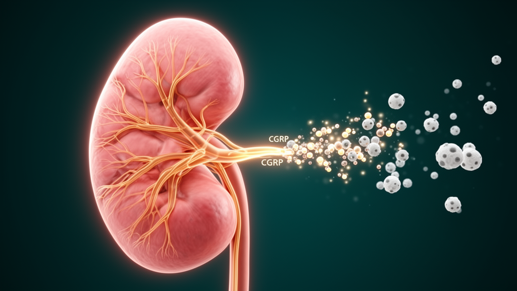
Sensory nerves that normally signal pain turn out to be unexpected guardians of the kidney, according to a new study in Cell Research. Using whole-organ tissue clearing and three-dimensional imaging, researchers mapped an extensive network of nociceptive sensory fibers threading through the renal parenchyma, far beyond the renal pelvis where afferent nerves were long thought to be confined. When those TRPV1-positive pain neurons were removed, mice suffered dramatically worse kidney injury after ischemia-reperfusion, and when the neurons were chemically activated, the injury was blunted. The work defines a dorsal root ganglion-to-kidney neuroimmune circuit and points to the neuropeptide CGRP as a potential therapeutic target for acute kidney injury and its progression to chronic kidney disease.
Acute kidney injury affects more than 13.3 million people worldwide each year, prolongs hospital stays, and carries mortality ranging from roughly 10 percent to 80 percent depending on the population and the severity of injury. Survivors, particularly older adults and people with diabetes or hypertension, face elevated risks of chronic kidney disease. No pharmacological therapy has yet been approved to prevent the condition or speed recovery, which makes the identification of a protective neural pathway especially significant.
The team, led by Lei Wang and Xin Ma of the Chinese PLA General Hospital, applied the SHANEL clearing method to whole mouse kidneys and reconstructed the three-dimensional architecture of CGRP-positive sensory fibers with light-sheet microscopy. To confirm the imaging genetically, they crossed TRPV1-Cre mice with fluorescent reporter lines and used CUBIC clearing to visualize labeled fibers within the kidney. Retrograde tracing with pseudorabies virus and anterograde tracing with a viral vector converging on the same answer showed that each kidney receives its nociceptive supply predominantly from the ipsilateral T10 through L2 dorsal root ganglia, with little contribution from the vagal sensory ganglia.
Functional experiments established that this innervation is not a passive wiring diagram. Three independent depletion strategies, genetic ablation with diphtheria toxin in Trpv1-Cre mice, systemic chemical ablation with the TRPV1 agonist resiniferatoxin, and localized capsaicin treatment around the renal artery, each worsened outcomes after bilateral ischemia-reperfusion. Ablated animals showed higher serum creatinine and urea on day two, more tubular necrosis, dilation and cast formation, and by day twenty-eight increased interstitial collagen deposition indicating progressive fibrosis. Conversely, chemogenetic activation of T10-L2 nociceptors using a DREADD receptor improved both acute and chronic outcomes. The authors note that all formal mechanistic experiments were conducted in female mice, because exploratory studies in males produced high mortality that left too few survivors for adequately powered analysis, so the conclusions are restricted to females.
Among the neuropeptides the researchers quantified in kidney tissue, only CGRP, the calcitonin gene-related peptide, was abundant at baseline and rose after injury, and its expression increased in the cell bodies of kidney-innervating dorsal root ganglion neurons. Mice lacking CALCA-derived alpha-CGRP developed more severe acute and chronic injury after ischemia-reperfusion, while systemic CGRP supplementation beginning two days before surgery and continuing for two weeks protected wild-type mice and rescued the exaggerated injury caused by sensory denervation. Together these experiments identified CGRP as the key mediator linking nociceptors to kidney protection.
The cellular target turned out to be the innate immune system. Flow cytometry of renal leukocytes showed that sensory denervation specifically increased infiltration of CD11b-high monocytes and macrophages and neutrophils, without changing dendritic cells, B cells, or T cells. To test whether CGRP acts on monocytes and macrophages, the team generated mice with myeloid-specific deletion of Calcrl, which encodes a component of the CALCRL-RAMP1 CGRP receptor complex. These mice phenocopied sensory ablation, with amplified myeloid infiltration and worse kidney injury, and exogenous CGRP no longer reduced leukocyte infiltration or protected the kidney in them. Reducing monocytes and macrophages with a CSF1R inhibitor abolished the excess inflammation and injury, confirming these cells as the principal effectors.
Transcriptomic analysis of sorted renal monocytes and macrophages revealed the mechanism. Without CGRP signaling, the cells upregulated TNF, Toll-like receptor and NF-kappa-B pathway modules along with pro-inflammatory cytokine genes such as Tnf and Il1b and chemokine genes including Cxcl2 and Ccl3, with corresponding increases in protein measured in whole-kidney lysates. In cultured bone-marrow-derived macrophages, CGRP suppressed the response to lipopolysaccharide, but this suppression failed in receptor-deficient cells and was restored by forskolin, an adenylyl cyclase activator that bypasses the receptor. Giving forskolin to receptor-deficient mice in vivo normalized cytokine and chemokine production, reduced monocyte and neutrophil influx, and improved both acute and chronic kidney outcomes, placing the cAMP-PKA pathway downstream of CALCRL-RAMP1 in this axis.
The most clinically striking finding concerns vulnerable populations. In a biospecimen cohort of forty-eight nephrectomy samples from macroscopically normal cortical tissue, renal CGRP levels were significantly lower in kidneys from elderly patients and from patients with diabetes than in young and middle-aged reference patients, while hypertensive samples showed no significant difference. Matching mouse models, streptozotocin and high-fat diet-induced diabetes and natural aging at twenty months, recapitulated the reduced baseline CGRP and the blunted rise after injury, with statistical analysis confirming significant interactions between diabetes or age and ischemic injury.
When diabetic and aged mice were subjected to ischemia-reperfusion, CGRP supplementation reduced TNF, IL-1beta, CXCL2 and CCL3, curtailed infiltration of neutrophils, monocytes and macrophages, and improved functional and histological outcomes at both acute and chronic time points. The protective effects were more pronounced in these high-risk models than in young healthy animals, suggesting that impaired CGRP signaling contributes to the heightened susceptibility of aged and diabetic kidneys and that restoring the pathway could be most valuable precisely where risk is greatest.
The authors are careful about the limits of the work. The upstream signals that activate renal nociceptors and trigger CGRP release remain unidentified, the central projections of the fibers were not mapped, interactions with sympathetic outflow were not fully delineated, and TRPV1-lineage tools label only a subset of nociceptors. Whether the pathway behaves similarly in males is unknown. Even so, the study extends the emerging framework of nociceptor-mediated immune regulation to the kidney, establishes the anatomical and molecular basis of a sensory protective circuit, and nominates CGRP signaling through CALCRL-RAMP1 and cAMP-PKA in myeloid cells as a plausible strategy for treating acute kidney injury and halting its slide toward chronic kidney disease.
Subject of Research: A dorsal root ganglion-kidney neuroimmune circuit in which sensory neuron-derived CGRP protects against ischemia-reperfusion kidney injury and fibrosis via myeloid cell signaling
Article Title: Sensory neuron-derived CGRP ameliorates kidney injury and fibrosis via monocytes and macrophages
Article References: Wang, L., Li, H., Feng, J., Chen, J., Jia, T., Zhou, T., Wang, L., Tang, L., Zhang, X., & Ma, X. (2026). Sensory neuron-derived CGRP ameliorates kidney injury and fibrosis via monocytes and macrophages. Cell Research. https://doi.org/10.1038/s41422-026-01308-7
Image Credits: AI Generated
DOI: 10.1038/s41422-026-01308-7
Keywords: acute kidney injury, chronic kidney disease, nociceptors, CGRP, neuroimmune crosstalk, macrophages, ischemia-reperfusion, fibrosis, dorsal root ganglia, diabetes, aging, cAMP-PKA pathway
Cite Scienmag News
Cassandra Pierce. (October 23, 2026). Pain-Sensing Nerves Protect the Kidney Through a CGRP Immune Circuit. Scienmag. https://scienmag.com/pain-sensing-nerves-protect-the-kidney-through-a-cgrp-immune-circuit/
Cassandra Pierce. "Pain-Sensing Nerves Protect the Kidney Through a CGRP Immune Circuit." Scienmag, 23 October 2026, https://scienmag.com/pain-sensing-nerves-protect-the-kidney-through-a-cgrp-immune-circuit/. Accessed 23 October 2026.
Cassandra Pierce. "Pain-Sensing Nerves Protect the Kidney Through a CGRP Immune Circuit." Scienmag. October 23, 2026. https://scienmag.com/pain-sensing-nerves-protect-the-kidney-through-a-cgrp-immune-circuit/


