


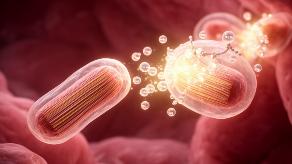
Cardiomyocytes, the contractile cells of the heart, do more than pump blood during embryonic development. According to a study published in Nature Cardiovascular Research, these cells secrete transforming growth factor β (TGFβ) ligands that orchestrate the maturation of the entire heart, including the cells' own final identity. When researchers deleted all three TGFβ ligand genes, Tgfb1, Tgfb2 and Tgfb3, specifically in developing mouse cardiomyocytes, the animals developed cardiac dysfunction by six weeks of age, accompanied by altered fibroblast activity, abnormal extracellular matrix properties, and changes in endothelial and immune cell populations.
The most striking finding emerged from gene profiling of the mutant hearts. Beginning at embryonic day 17.5, cardiomyocytes lacking TGFβ ligands started expressing skeletal muscle genes, a program that should remain silent in heart muscle. This ectopic expression indicates that the cells failed to complete their normal maturation into fully committed adult myocytes.
TGFβ ligands are secreted factors sequestered in the extracellular matrix, and their role in heart maturation had previously been unclear. To determine how cardiomyocyte-derived TGFβ exerts its effects, the team ran a series of genetic tests. Deleting the TGFβ receptors Tgfbr1 and Tgfbr2 in cardiomyocytes, or removing the downstream signaling mediators Smad2 and Smad3, did not reproduce the defects seen when the ligands themselves were deleted.
This indicates that the TGFβ produced by heart muscle cells does not act back on the cardiomyocytes through canonical signaling; instead, it signals to non-myocyte neighbors. Those neighbors include cardiac fibroblasts, which the study found to be improperly matured in the mutant hearts, along with a disorganized extracellular matrix. The researchers then tested whether the problem was simply mechanical, arising from a defective matrix environment.
Mice lacking the matrix components collagen I and VI (Col1a2−/−;Col6a2−/−), which have reduced matrix stiffness, did not show the ectopic skeletal muscle gene program. Similarly, cardiomyocyte-specific deletion of Itgb1, which impairs the cells' ability to sense matrix mechanics, and loss of cardiac fibroblasts in Tcf21−/− embryos each failed to reproduce the phenotype. Together, these experiments suggest that embryonic cardiomyocyte-derived TGFβ establishes a signaling environment through non-myocytes that directs cardiomyocyte maturation and identity, a mechanism distinct from simple matrix stiffness or mechanosensing.
Among the three ligands, Tgfb1 alone appeared capable of driving cardiomyocyte maturation, while Tgfb2 and Tgfb3 did not. Single-nucleus sequencing of the mutant hearts further revealed reduced attachment complexes, consistent with the observed defects in cell-matrix interactions. The mutant hearts at six weeks of age also showed fibrosis and deficient fibroblast maturation, painting a picture of a heart that had not properly completed its developmental program.
Interestingly, the researchers found no changes in epicardial epithelial-to-mesenchymal transition or in the migration of epicardial-derived cells, indicating that the developmental defects were selective rather than global, and that the TGFβ signal acts on specific downstream cellular programs during maturation. The gene profiling experiments traced the earliest molecular defect to E17.5, when ectopic skeletal muscle gene expression first appeared in cardiomyocytes lacking the ligands. This timing allowed the team to place the requirement for cardiomyocyte-derived TGFβ in the late embryonic period, before the heart has fully assumed its postnatal workload.
The reprogramming of fibroblasts in the mutant hearts was associated with the aberrant cardiomyocyte differentiation, reinforcing the paracrine nature of the circuit. Because the study was conducted entirely in mice using cardiomyocyte-specific genetic deletions, its findings describe developmental mechanisms in an animal model rather than clinical outcomes in humans. Nevertheless, the work identifies a previously unrecognized paracrine circuit in which heart muscle cells shape their own destiny by instructing surrounding cells, and it maps the timing, ligand specificity and cellular mediators of that circuit with genetic precision.
The broader significance lies in redefining what a cardiomyocyte does during development. Rather than being passive recipients of maturation cues from their environment, these cells appear to be active participants, secreting TGFβ ligands that are stored in the surrounding matrix and that instruct fibroblasts and other non-myocytes to create conditions permissive for proper myocyte identity. Disruption of this loop leads to a cascade of secondary defects, including endothelial and immune changes, abnormal matrix properties, and ultimately cardiac dysfunction in young adult animals.
By systematically ruling out cell-autonomous receptor signaling, matrix stiffness, mechanosensing and fibroblast absence as sufficient explanations for the phenotype, the study narrows the search for the true mediators of the maturation signal.
Subject of Research: The role of cardiomyocyte-derived TGFβ signaling in mouse heart maturation and myocyte identity
Article Title: Cardiomyocyte-expressed TGFβ programs early heart maturation and adult myocyte identity
Article References: Minerath, R. A., Kasam, R. K., Swoboda, C. O., Prasad, V., Grimes, K. M., Blair, N. S., Khalil, H., Alfieri, C. M., Eads, L., Saviola, A. J., Azhar, M., Miao, L., Wu, M., Tallquist, M., Hansen, K. C., Weirauch, M. T., Yutzey, K. E., Millay, D. P., & Molkentin, J. D. (2026). Cardiomyocyte-expressed TGFβ programs early heart maturation and adult myocyte identity. Nature Cardiovascular Research. https://doi.org/10.1038/s44161-026-00892-6
Image Credits: AI Generated
DOI: 10.1038/s44161-026-00892-6
Keywords: TGFβ, cardiomyocytes, heart development, cardiac fibroblasts, extracellular matrix, mouse genetics, maturation, skeletal muscle genes, Smad signaling, single-nucleus sequencing, paracrine signaling, Nature Cardiovascular Research
Cite Scienmag News
Juliet Wilcox. (October 23, 2026). Heart Muscle Cells Signal Their Own Maturation Through TGFβ, Mouse Study Finds. Scienmag. https://scienmag.com/heart-muscle-cells-signal-their-own-maturation-through-tgf%ce%b2-mouse-study-finds/
Juliet Wilcox. "Heart Muscle Cells Signal Their Own Maturation Through TGFβ, Mouse Study Finds." Scienmag, 23 October 2026, https://scienmag.com/heart-muscle-cells-signal-their-own-maturation-through-tgf%ce%b2-mouse-study-finds/. Accessed 23 October 2026.
Juliet Wilcox. "Heart Muscle Cells Signal Their Own Maturation Through TGFβ, Mouse Study Finds." Scienmag. October 23, 2026. https://scienmag.com/heart-muscle-cells-signal-their-own-maturation-through-tgf%ce%b2-mouse-study-finds/


