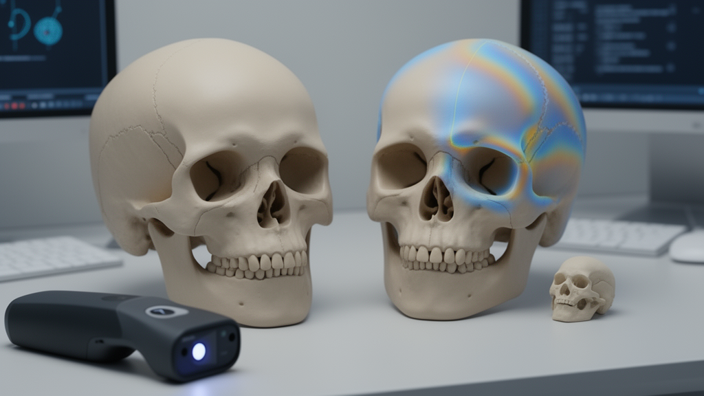
A computational workflow combining structured-light scanning, image registration and shape analysis enabled researchers to quantify changes to an infant’s skull during corrective surgery for craniosynostosis. The method was applied to two cases: one infant with trigonocephaly and one with Saethre–Chotzen syndrome. In both patients, the analyses indicated frontal advancement in the anterior and lateral directions, providing a preliminary clinical demonstration rather than broad validation across a patient cohort.
The study, published in the International Journal of Computer Assisted Radiology and Surgery , focused on quantitative assessment during fronto-orbital advancement and remodelling. The investigators noted that intraoperative evaluation currently relies primarily on surgeons’ judgment and clinical experience, and they tested whether a three-dimensional scan could support a more detailed morphological record. The researchers built the workflow in three stages.
They first computed cephalometric measurements, including the interfrontal angle and cranial index, from the intraoperative three-dimensional surface scan obtained with a structured-light handheld scanner. They then used a coarse-to-fine registration method to align the preoperative and intraoperative surfaces, enabling the non-operated region to be superimposed as a reference. Finally, geometric morphometrics was used to examine differences in frontal shape, while colour maps represented intraoperative geometric changes across the scanned surface.
Alignment produced a root mean square error of 0.5–0.6 millimetres, which the investigators interpreted as indicating high registration accuracy. The cephalometric measurements and colour maps showed frontal advancement in both the anterior and lateral directions. The interfrontal angle increased in the trigonocephaly case, whereas the cranial index decreased in the Saethre–Chotzen syndrome case.
The supplied abstract does not report the numerical change in every measurement or the size and composition of the comparison group. Geometric morphometric analysis suggested that the central forehead remained relatively flat on the intraoperative three-dimensional surface. At the same time, the overall frontal morphology approached that of the control group.
The study therefore combined three forms of assessment: conventional cephalometric measurements, registration-based comparison of pre- and intraoperative surfaces, and geometric analysis of morphological variation. Together, they provided a quantitative description of where and how the frontal region had changed in the two operated infants. It was applied to only two patients, each with a different craniosynostosis diagnosis.
The reported registration error describes the accuracy of aligning the surfaces, but it does not by itself establish clinical benefit or show how the measurements would perform in a larger and more diverse surgical series. The abstract also does not report patient outcomes, operative complications, operating time or the effect of quantitative feedback on surgical decisions. Accordingly, the findings show that three-dimensional scanning and computational analysis can document intraoperative skull remodelling in individual craniosynostosis cases.
They do not demonstrate that quantitative assessment improves clinical results, reduces complications or shortens surgery. The investigators proposed that the framework could be integrated into routine surgical workflows, but that possibility remains a stated potential rather than a validated outcome of the two-patient study. The method links an intraoperative scan to preoperative anatomy through registration, then tests for changes using both named cephalometric indicators and overall shape analysis.
Further clinical evaluation would be needed to establish consistency, usefulness and the conditions under which this quantitative approach could add value to conventional intraoperative assessment in craniosynostosis treatment.
Subject of Research: Intraoperative 3D morphometric assessment of skull remodelling during craniosynostosis surgery.
Article Title: Quantitative intraoperative assessment of fronto-orbital advancement and remodelling in craniosynostosis
Article References: Shui, W., Vakharia, V., de Gea Rico, A., Van Carlen, M., Liang, C., Moazen, M., Khonsari, R. H., Hennedige, A., Sinha, A., Parks, C., & Duncan, C. (2026). Quantitative intraoperative assessment of fronto-orbital advancement and remodelling in craniosynostosis. International Journal of Computer Assisted Radiology and Surgery. https://doi.org/10.1007/s11548-026-03807-9
Image Credits: AI Generated
DOI: 10.1007/s11548-026-03807-9
Keywords: craniosynostosis, 3D surface scanning, intraoperative imaging, skull remodelling, image registration, cephalometric analysis, geometric morphometrics, trigonocephaly, Saethre–Chotzen syndrome, computer-assisted surgery, Quantitative, intraoperative
Cite Scienmag News
Ophelia Keating. (October 22, 2026). 3D scan method quantifies skull remodelling in two craniosynostosis cases. Scienmag. https://scienmag.com/3d-scan-method-quantifies-skull-remodelling-in-two-craniosynostosis-cases/
Ophelia Keating. "3D scan method quantifies skull remodelling in two craniosynostosis cases." Scienmag, 22 October 2026, https://scienmag.com/3d-scan-method-quantifies-skull-remodelling-in-two-craniosynostosis-cases/. Accessed 22 October 2026.
Ophelia Keating. "3D scan method quantifies skull remodelling in two craniosynostosis cases." Scienmag. October 22, 2026. https://scienmag.com/3d-scan-method-quantifies-skull-remodelling-in-two-craniosynostosis-cases/