


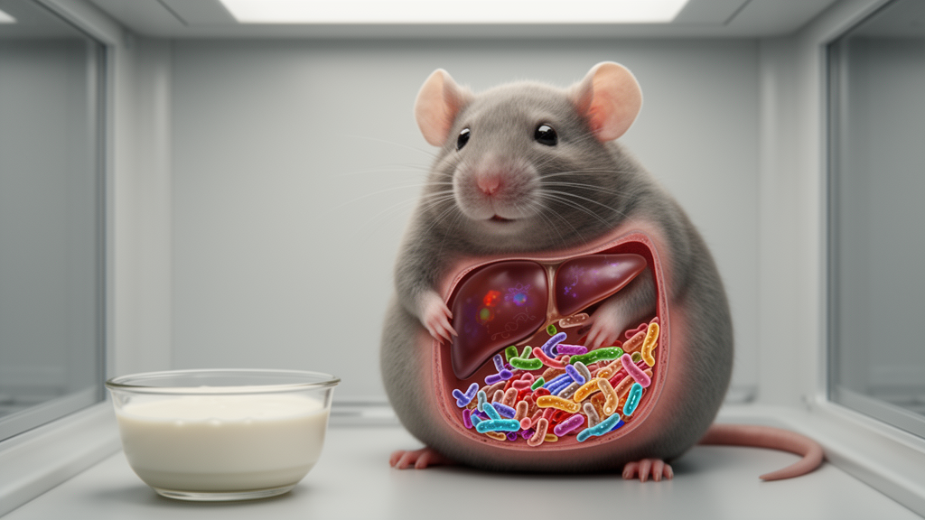
Daily camel milk reduced body weight, fat accumulation, blood lipid levels and liver fat in male mice fed a high-fat diet, according to a multi-omics study that associated these metabolic improvements with partial changes in the gut microbiota, short-chain fatty acid concentrations and hepatic gene expression. The experiment also found that camel-milk-treated mice had an intermediate hepatic metabolome and transcriptome between normal-diet controls and untreated high-fat-diet animals, indicating that the intervention did not fully reverse diet-induced metabolic disruption. These results establish a preclinical association, not a clinical treatment for obesity.
The researchers used five-week-old male C57BL/6J mice, which were acclimatized for one week and then assigned by computer-generated randomization, with body-weight stratification, to four groups of eight. One group received a normal diet, while the other three received a high-fat diet. After 10 weeks on the diets, one high-fat-diet group received saline by gavage, another received the anti-obesity drug orlistat, and the third received raw camel milk at 10 millilitres per kilogram of body weight daily for seven weeks.
The camel milk was collected during mid-lactation in Inner Mongolia, kept frozen at minus 80 degrees Celsius, thawed before administration and delivered without pasteurization. Investigators conducting biochemical assays, sequencing-library preparation and bioinformatics analyses were blinded to group assignment. Compared with untreated high-fat-diet mice, those receiving camel milk weighed significantly less at weeks 13 through 16 and had lighter inguinal and epididymal white adipose tissue.
Their adipocytes also occupied less area on haematoxylin and eosin-stained sections, a histological measure of reduced cellular enlargement. Serum triglycerides and low-density lipoprotein cholesterol fell, while high-density lipoprotein cholesterol rose. Food intake was lower in the camel-milk group during weeks 14 and 15, a finding that may partly account for the weight difference and is one of the study’s central limitations.
Because researchers did not include an isocaloric pair-fed control, they could not separate effects of reduced food consumption from effects potentially attributable to specific components of camel milk. The metabolic effects were not uniform. Camel milk significantly reduced serum alanine aminotransferase and attenuated the lipid droplets seen in liver sections, both consistent with less hepatic steatosis, but its reduction in liver weight was not statistically significant.
Fasting blood glucose, fasting insulin and the homeostasis model assessment of insulin resistance did not differ significantly among the four groups. In the oral glucose tolerance test, camel-milk-treated mice showed an improved glucose-response profile, although the clearest statistical glucose reduction was reported for orlistat. Insulin sensitivity improved significantly with orlistat, whereas the improvement in the camel-milk group was not significant.
Random blood glucose was also lower with orlistat but not significantly lower with camel milk. The result therefore appears to involve improved glucose tolerance alongside improved adiposity and blood lipids, rather than a demonstrated correction of every measure of metabolic dysfunction. Sequencing of bacterial 16S rRNA genes from colonic contents showed that the high-fat diet separated the microbiome from the normal-diet group and that camel milk shifted it only partly back.
Principal coordinate analysis placed the camel-milk group between the normal-control and high-fat-diet clusters, with the dominant separation along the first axis accounting for 68.43% of the variation. At the phylum level, camel milk increased Bacteroidota and reduced Pseudomonadota and Deferribacterota relative abundance compared with the untreated high-fat-diet group. It also suppressed Mucispirillum, Escherichia-Shigella and Faecalitalea, partially restored Bacteroides and increased Parabacteroides.
The unclassified Eubacterium coprostanoligenes group became particularly abundant after treatment. These are compositional findings: they show that taxa changed alongside improved metabolic phenotypes, but they do not demonstrate that any organism caused the improvements. Targeted measurements in colonic contents found that the high-fat diet lowered concentrations of several short-chain fatty acids, including acetic, butyric and propionic acids.
Camel milk raised all eight measured compounds relative to the untreated high-fat-diet group, with acetic and propionic acid returning to levels comparable to those of normal-diet controls. The restoration of these bacterial metabolites is consistent with a mechanism involving the gut-liver axis, through which intestinal organisms and their products could influence liver metabolism. Nevertheless, the short-chain-fatty-acid analysis used only three biological replicates per group, and the researchers emphasized the potential variability of these measurements.
The study also did not identify which dietary substrates or microbial pathways produced the changes, so the milk cannot yet be classified as a prebiotic on the basis of these data alone. Untargeted liver metabolomics revealed a broad but incomplete remodeling of hepatic chemistry. The camel-milk samples clustered between normal controls and high-fat-diet animals in principal component analysis.
In positive-ion mode, researchers identified 443 differential metabolites between normal controls and untreated high-fat-diet mice, compared with 170 between camel-milk and untreated mice; the corresponding negative-ion counts were 244 and 158. Camel milk reversed the high-fat-diet-associated elevations of sphingosine and the sphingomyelin SM(d18:0/16:1(9Z)). Enrichment analysis implicated lipid and sphingolipid signaling, arachidonic acid metabolism, amino-acid pathways, glutathione metabolism, oxidative stress and inflammatory signaling.
These findings identify differential metabolites and enriched metabolic pathways, but the metabolomic analysis alone cannot establish which biochemical reactions caused the metabolic phenotypes or demonstrate changes in metabolic flux. RNA sequencing likewise showed partial correction at the transcriptional level. Compared with untreated high-fat-diet mice, camel-milk-treated livers had 613 differentially expressed genes, including 181 upregulated and 432 downregulated genes; 271 of these genes had also differed between normal controls and high-fat-diet animals.
Changes involved arachidonic acid metabolism, fatty-acid elongation, unsaturated-fatty-acid biosynthesis, glycerolipid metabolism and bile secretion. Among the changes were lower expression of several Cyp4a genes, multiple Acot genes and the glycerolipid genes Aldh3a2, Mgll and Mogat1. The bile-transport gene Abcg5 was upregulated, whereas several other bile-related transcripts changed in the opposite direction.
These findings indicate transcriptional reprogramming, not demonstrated changes in enzyme activity, metabolic flux, bile secretion or reverse cholesterol transport. The researchers did not directly measure 20-hydroxyeicosatetraenoic acid, the relevant output of Cyp4a enzymes, and did not validate the selected transcripts at the protein level. Spearman correlations connected several microbial taxa with metabolic and molecular measurements.
The unclassified Eubacterium coprostanoligenes group was negatively associated with epididymal fat weight at r = −0.846, inguinal fat weight at r = −0.818, triglycerides at r = −0.673 and the oral-glucose-test area under the curve at r = −0.580. Faecalitalea showed opposite associations with some fat and lipid measures. The Eubacterium group was positively associated with metabolites linked to glutathione, amino-acid and carbohydrate metabolism and negatively associated with sphingolipids and unsaturated fatty acids, while Escherichia-Shigella displayed largely opposing patterns.
It also correlated with lipid-metabolism genes, including Cyp2c70, several Cyp4a and Acot genes, Aldh3a2 and Ugt1a7c. Such cross-omics associations strengthened the proposed link between the altered microbiome and hepatic lipid regulation, but they remain correlational and cannot establish direction of effect or mediation. Several further boundaries constrain interpretation.
The experiment included only male mice, so sex-specific responses remain unknown. It lacked a comparison with conventional bovine or goat milk and therefore cannot show that the effects were unique to camel milk. Raw milk was used to preserve potentially heat-sensitive constituents such as lactoferrin, lysozyme, immunoglobulins and insulin-like proteins, but trace microbial products or enzymes could also have contributed.
The multi-omics components used different sample sizes, including three replicates for short-chain fatty acids and transcriptomics, and the study tested whole milk rather than isolated molecules. The defensible conclusion is that camel milk produced measurable metabolic and molecular changes in this mouse model, with a possible role for microbial and short-chain-fatty-acid changes in liver lipid remodeling. Fecal microbiota transfer, microbial depletion, pair-feeding, other-milk controls and eventual human studies would be needed to determine whether camel milk itself has a reproducible, causal and clinically useful effect.
Subject of Research: Camel milk effects on gut microbiota and hepatic lipid metabolism in high-fat-diet-fed mice
Article Title: Camel milk is associated with ameliorated high-fat diet-induced obesity and metabolic disorders via modulation of gut microbiota and remodeling of hepatic lipid metabolism: a multi-omics analysis
Article References: Yang, B., Wang, J., Pan, W., Liang, H., & Zhao, H. (2026). Camel milk is associated with ameliorated high-fat diet-induced obesity and metabolic disorders via modulation of gut microbiota and remodeling of hepatic lipid metabolism: a multi-omics analysis. Frontiers in Nutrition, 13, Article 1913436. https://doi.org/10.3389/fnut.2026.1913436
Image Credits: AI Generated
DOI: 10.3389/fnut.2026.1913436
Keywords: camel milk, gut microbiome, gut-liver axis, diet-induced obesity, short-chain fatty acids, hepatic steatosis, lipid metabolism, multi-omics, metabolic disorders, mouse model, Camel, milk
Cite Scienmag News
Daisy Hatcher. (October 22, 2026). Camel milk alters gut bacteria and liver metabolism in obese mice. Scienmag. https://scienmag.com/camel-milk-alters-gut-bacteria-and-liver-metabolism-in-obese-mice/
Daisy Hatcher. "Camel milk alters gut bacteria and liver metabolism in obese mice." Scienmag, 22 October 2026, https://scienmag.com/camel-milk-alters-gut-bacteria-and-liver-metabolism-in-obese-mice/. Accessed 22 October 2026.
Daisy Hatcher. "Camel milk alters gut bacteria and liver metabolism in obese mice." Scienmag. October 22, 2026. https://scienmag.com/camel-milk-alters-gut-bacteria-and-liver-metabolism-in-obese-mice/


