


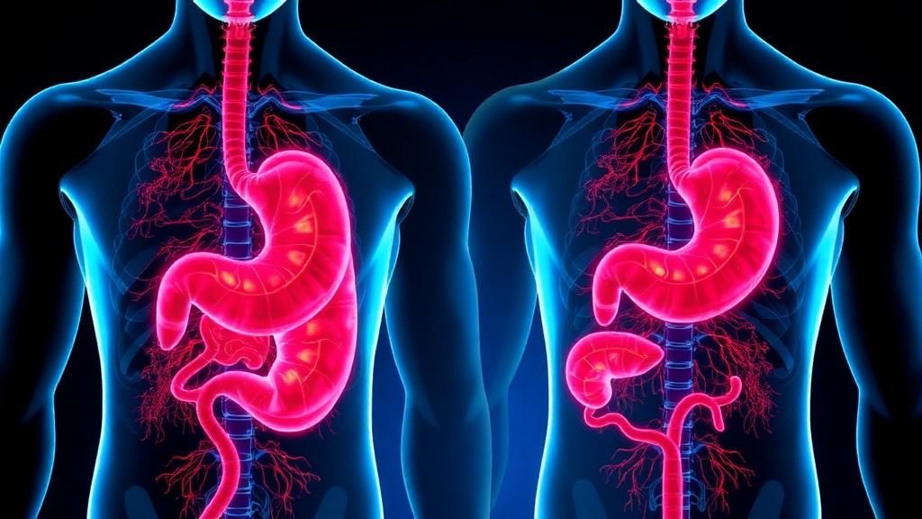
A 2026 narrative review proposes that diabetes mellitus and atrophic gastritis may be linked by a bidirectional feedback loop: persistent hyperglycemia can promote gastric mucosal injury, while the loss of gastric function in atrophic gastritis may impair glucose metabolism. The authors bring together animal experiments, cross-sectional observations, retrospective studies and genetic associations, but emphasize that most of the proposed causal links have not been tested in large prospective cohorts or interventional trials involving people with both diseases. The central model is therefore a hypothesis for investigation rather than an established clinical mechanism, and its strongest support comes from biological convergence among oxidative stress, inflammation, impaired mitochondrial quality control, nutrient deficiency and altered gastrointestinal hormones.
On the diabetes-to-stomach side of the model, chronic hyperglycemia is presented as the initiating pressure. Excess glucose overloads mitochondrial electron transport, increases reactive oxygen species and activates pathways including NADPH oxidase. In diabetic animals, the review reports higher concentrations of lipid peroxidation products such as MDA and 4-HNE in gastric mucosa, reduced antioxidant enzyme activity, activation of the NF-κB inflammatory axis, an unfavorable balance between the anti-apoptotic protein Bcl-2 and pro-apoptotic Bax, and activation of caspase-9 and caspase-3. The resulting epithelial apoptosis, impaired cell proliferation and thinned lamina propria provide a plausible route from hyperglycemia to gland loss and mucosal atrophy. This evidence comes primarily from experimental models and does not establish the same sequence in human diabetes.
Advanced glycation end products, or AGEs, add another proposed route. High glucose promotes non-enzymatic glycation of long-lived matrix proteins including collagen and laminin, altering their cross-linking and physical properties. When AGEs engage the receptor for AGEs, or RAGE, they can activate NF-κB and MAPK signaling, stimulate NADPH oxidase and promote further reactive oxygen species. Animal studies summarized in the review associate elevated AGEs with structural disorders, while experimental findings connect RAGE signaling to impaired fibroblast function, reduced collagen synthesis and delayed healing. AGE cross-linking may also make the mucosa less compliant and more vulnerable to acid, bile and Helicobacter pylori. The authors describe this as a positive feedback loop in which oxidative stress and glycation reinforce one another, but they acknowledge that direct longitudinal evidence in patients remains limited.
Diabetes may also compromise the stomach’s blood supply and its capacity to repair. Diabetic microangiopathy affects the small vessels beneath the gastric mucosa, producing endothelial swelling, luminal narrowing, reduced perfusion density and chronic ischemia. The review cites measurements showing lower mucosal blood flow in people with diabetes than in healthy controls and patients with chronic superficial gastritis. Reduced nitric oxide bioavailability, increased asymmetric dimethylarginine and elevated von Willebrand factor can further disturb microcirculation and microthrombosis. Prolonged hypoxia stabilizes HIF-1α, which may initially support adaptation but, when chronically imbalanced, can sustain inflammatory signaling, reduce tight-junction proteins and hinder epithelial regeneration. Deficits in prostaglandin E2, epidermal growth factor and TGF-β signaling are also described as weakening mucus and bicarbonate protection and slowing the reconstruction of damaged epithelium.
Neurological complications add a gastric-motility component. Diabetic autonomic neuropathy can reduce vagal stimulation of acid-secreting parietal cells, disturb the balance between gastrin-secreting G cells and somatostatin-secreting D cells, and impair the protective role of ghrelin. Diabetic gastroparesis is characterized in the review by weak antral contractions, increased pyloric resistance and delayed emptying of solid food. Retention and reflux of bile- and enzyme-rich duodenal contents can damage epithelial tight junctions and cell membranes. Loss or dysfunction of interstitial cells of Cajal, the stomach’s electrical pacemaker cells, further disrupts contractions, and experimental work links oxidative stress, microvascular injury, pro-inflammatory macrophages and altered autophagy to their decline. Together, delayed emptying, bile reflux, mucosal injury, cellular loss and worsening motility are proposed to form a self-reinforcing sequence that can progress from chronic inflammation to gland atrophy and intestinal metaplasia.
Helicobacter pylori provides a potential meeting point between hyperglycemia, infection and atrophy. The review describes altered glycosylation of gastric mucins in a high-glucose environment, which may expose structures that improve bacterial adhesion, together with reduced acidity that weakens the stomach’s chemical defense. Diabetes-associated impairment of neutrophil chemotaxis, phagocytosis and intracellular killing may make early clearance less effective, while macrophage responses become more strongly skewed toward inflammation. H. pylori can also induce the immunosuppressive cytokine IL-37 through receptors including TLR2, TLR4 and NOD1. Its virulence factors, notably CagA from cag-positive strains, can amplify NADPH oxidase activity, oxidative damage and NF-κB signaling, potentially accelerating the sequence from gastritis to atrophy and metaplasia. These observations suggest that infection, immune dysfunction and high glucose may cooperate, but they do not show that every patient with diabetes follows this course or that eradication alone prevents atrophy.
In the reverse direction, atrophic gastritis may disturb glucose metabolism through several partially overlapping mechanisms. Loss of parietal cells reduces gastric acid and intrinsic factor, impairing the release and terminal-ileal absorption of vitamin B12 and reducing the conversion of dietary iron to a more soluble form. B12 deficiency can lead to hyperhomocysteinemia, which has been associated with oxidative stress, endothelial injury and insulin resistance. Preclinical evidence also links homocysteine to endoplasmic-reticulum stress, mitochondrial dysfunction and apoptosis in pancreatic beta cells. Iron deficiency may restrict mitochondrial complex IV activity and ATP production in insulin-sensitive tissues, potentially worsening insulin signaling and glucose-stimulated insulin secretion. These pathways are biologically coherent, but the review notes that much of the evidence is preclinical and that trials demonstrating improved glycemic control after correction of atrophic-gastritis-related deficiencies are scarce.
Changes in the gastric endocrine network offer a second potential route. Loss of acid removes negative feedback on antral G cells, producing marked hypergastrinemia, while impaired D-cell function may weaken somatostatin inhibition. Although moderate gastrin elevations can have metabolic or trophic effects under physiological conditions, the review proposes that sustained supraphysiological stimulation of CCK-B receptors on beta cells may desensitize insulin secretion, deplete functional reserves and interfere with beta-cell transcription factors. Atrophic gastritis affecting the gastric body and fundus can also destroy X/A-like cells, the main source of ghrelin, altering meal-related hunger signals and potentially removing ghrelin’s inhibitory influence on basal insulin secretion. Because ghrelin also affects skeletal-muscle mitochondrial function and inflammation, its loss may intersect with diabetic sarcopenia and insulin resistance. These consequences remain mechanistically inferred rather than demonstrated by prospective hormone-intervention trials.
Low gastric acidity can also permit small intestinal bacterial overgrowth, altering microbial metabolites and the gut-liver-pancreas axis. Lipopolysaccharide from Gram-negative bacteria may enter portal circulation, activate TLR4 and inflammatory pathways such as JNK and IKK/NF-κB, and contribute to systemic insulin resistance. Changes in short-chain fatty acids and bile-acid metabolism may affect intestinal barrier integrity and the incretin hormones GLP-1 and GIP. The review reports associations between overgrowth, dysbiosis, irregular glucose regulation and reduced post-meal incretin responses, but notes that microbial and short-chain-fatty-acid responses can be complex and cannot be reduced to a single pathway. A major shared mechanism in the proposed cycle is disruption of the PINK1/Parkin mitophagy pathway: experimental findings associate high glucose and inflammation with reduced Parkin activity, damaged mitochondrial accumulation, impaired autophagic flux and apoptosis in both gastric epithelium and pancreatic beta cells, while leaked mitochondrial DNA can further activate STING and NF-κB signaling.
The review also identifies a shared immune and genetic background. HLA class II haplotypes such as DR3-DQ2 and DR4-DQ8 have been associated with susceptibility to both diseases, while anti-islet, anti-parietal-cell and intrinsic-factor antibodies occur in subsets of patients with diabetes. An imbalance between pro-inflammatory Th17 cells and regulatory T cells is another proposed link, with IL-17A activating NF-κB and STAT3-driven differentiation potentially contributing to injury of both gastric parietal cells and pancreatic beta cells. Yet the authors are explicit that none of the principal connecting nodes has been validated in a large prospective interventional study of comorbid patients. They call for longitudinal cohorts that repeatedly measure glucose control, gastroscopy, mucosal histology and biomarkers; dual-disease animal models and patient-derived gastric organoids; and simultaneous profiling of gastric and metabolic pathways. The review’s proposed antioxidant, hormonal, microecological and autophagy-based interventions are consequently early-stage research directions, not proven treatments for breaking the diabetes–gastritis cycle.
Subject of Research: Proposed bidirectional mechanisms linking diabetes mellitus and atrophic gastritis
Article Title: Diabetes mellitus and atrophic gastritis: progress in the study of interactive pathogenesis
Article References: Zhu, Z., Wang, L., Zhou, T., Che, Y., Shi, Y., Song, J., Zhang, Z., Xu, J., Tian, S., Qu, N., Lu, Y., Hao, Y., Zhang, J., Gao, H., & Lv, H. (2026). Diabetes mellitus and atrophic gastritis: progress in the study of interactive pathogenesis. Frontiers in Endocrinology, 17, Article 1955709. https://doi.org/10.3389/fendo.2026.1955709
Image Credits: AI Generated
DOI: 10.3389/fendo.2026.1955709
Keywords: diabetes mellitus, atrophic gastritis, hyperglycemia, gastric mucosa, oxidative stress, gut microbiome, insulin resistance, vitamin B12, gastroparesis, Helicobacter pylori, Diabetes, mellitus
Cite Scienmag News
Daisy Hatcher. (October 21, 2026). Review links diabetes and atrophic gastritis in a proposed feedback loop. Scienmag. https://scienmag.com/review-links-diabetes-and-atrophic-gastritis-in-a-proposed-feedback-loop/
Daisy Hatcher. "Review links diabetes and atrophic gastritis in a proposed feedback loop." Scienmag, 21 October 2026, https://scienmag.com/review-links-diabetes-and-atrophic-gastritis-in-a-proposed-feedback-loop/. Accessed 21 October 2026.
Daisy Hatcher. "Review links diabetes and atrophic gastritis in a proposed feedback loop." Scienmag. October 21, 2026. https://scienmag.com/review-links-diabetes-and-atrophic-gastritis-in-a-proposed-feedback-loop/


