


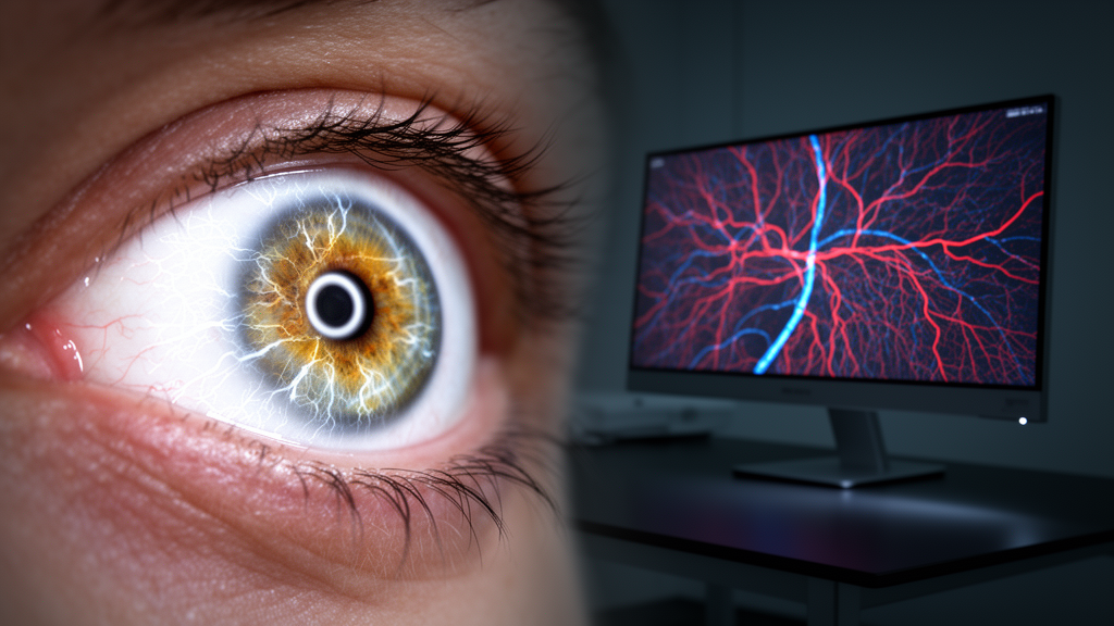
A 2026 structured narrative review concludes that strong performance on individual ophthalmic imaging datasets does not consistently produce reliable clinical measurements from artificial intelligence systems for vessel segmentation. The authors argue that clinically useful systems require validation across acquisition, anatomical preprocessing, segmentation, biomarker calculation and deployment.
The review covers retinal vascular trees in color fundus photography, scanning laser ophthalmoscopy and ultra-widefield imaging; capillary plexuses in optical coherence tomography angiography; structural OCT analysis of the choroid; macular neovascularization; corneal neovascularization; and conjunctival microvasculature. It is a purposive narrative synthesis rather than a systematic review or meta-analysis. The authors did not rerun algorithms or reanalyze metrics from raw predictions, and reported results are descriptive because datasets, populations, splits, annotations, thresholds and post-processing differ.
Vessels pose specific challenges for computational models: they have high aspect ratios, occupy a small fraction of many images, branch across several scales and may be only one or two pixels wide. A local break can disconnect a distal subtree while producing little change in a pixel-overlap measure such as Dice. Because background pixels dominate fundus images, overall accuracy can conceal poor sensitivity to fine terminal branches, low-contrast vessels and pathological networks. The review therefore considers multiscale feature pyramids, U-Net skip connections, deformable and directional operators, attention mechanisms, Transformers, state-space models, diffusion approaches and foundation models.
Architectural complexity alone did not emerge as a reliable predictor of usefulness. A Little W-Net study with 68,482 parameters reported Dice scores of 0.8279 on DRIVE, 0.8169 on CHASE_DB1 and 0.8103 on HRF. In a separate cross-dataset experiment without target-dataset retraining, the model reported Dice scores of 0.8282 on DRIVE and 0.3829 on UoA-DR. The latter result indicates substantial deterioration across datasets, although the studies used their own evaluation protocols and the review did not independently recompute the metrics.
OCT angiography introduces errors before vessel segmentation begins. It measures flow-related decorrelation rather than vessel walls, and en face slabs depend on automated structural-layer boundaries. Macular edema, drusen, high myopia, discontinuous segmentation and poor signal can displace those boundaries, while projection can duplicate superficial vessels in deeper slabs and motion can create breaks. Consequently, vessel density, skeleton density, foveal avascular zone area and fractal dimension reflect acquisition and processing as well as anatomy.
Several related tasks require different terminology and validation. Structural OCT choroidal vascularity index analysis divides choroidal tissue into luminal and stromal compartments rather than tracing a vessel tree. OCTA choriocapillaris analysis maps reduced flow signal, and repeatable thresholding methods can still yield measurements that are not interchangeable. Macular neovascularization is a pathological lesion-segmentation task, while anterior-segment hyperemia grading is a global redness assessment rather than vessel-pixel or centerline segmentation.
The review organizes evaluation at five levels: pixel overlap, centerline recovery, vascular topology, biomarker agreement and clinical actionability. Centerline distance, clDice, junction measures, connected-component errors and graph comparisons can assess thin-vessel localization and connectivity, but reference graph definitions and skeletonization remain variable. Biomarker validation should include bias, limits of agreement, intraclass correlation and repeatability. In an OCTA benchmark, U-Net and CS-Net each reported a Dice score of 0.89, while differences of up to 25% in vessel-density accuracy were reported across segmentation methods, illustrating why pixel overlap alone may not establish biomarker validity.
Physical calibration and cross-domain robustness are also central. Caliber, length and area require a calibrated lateral scale, while fundus and OCT systems often rely on model-eye assumptions influenced by axial length, refractive error and keratometry. Studies should state whether scaling occurs before or after segmentation, identify the formula and assumed model eye, and report results in pixels and physical units. Domain shifts may arise from camera spectrum, resolution, field of view, compression, pigmentation, disease mix, scan protocol, slab algorithm or annotation style, supporting external testing with untouched datasets, patient-level separation and patient-level confidence intervals.
The review’s proposed translation checklist covers acquisition controls, anatomical-preprocessing quality checks, uncertainty and failure-state reporting, DICOM and PACS interoperability, prospective impact studies and software-as-a-medical-device regulation. It presents a progression from technical validation to external retrospective testing, silent prospective deployment, workflow studies and management-impact evaluation, but does not claim that reviewed systems have completed those steps. Limitations include purposive selection, possible selection and publication bias, incomplete access to some reports, and results that cannot be directly compared when populations, splits, thresholds and annotations differ.
Subject of Research: A structured narrative review of artificial intelligence methods and validation requirements for ophthalmic vessel segmentation across multimodal imaging.
Article Title: Artificial intelligence for ophthalmic vessel segmentation: from multimodal imaging to clinical translation
Article References: Wu, L., Tang, J., & Zhao, S. (2026). Artificial intelligence for ophthalmic vessel segmentation: from multimodal imaging to clinical translation. Frontiers in Medicine, 13, Article 1912838. https://doi.org/10.3389/fmed.2026.1912838
Image Credits: AI Generated
DOI: 10.3389/fmed.2026.1912838
Keywords: ophthalmic vessel segmentation, artificial intelligence, retinal imaging, optical coherence tomography angiography, clinical validation, biomarkers, medical devices, image quality control, Artificial, intelligence, ophthalmic, vessel
Cite Scienmag News
Ophelia Keating. (October 21, 2026). Structured narrative review maps validation needs for ophthalmic vessel AI. Scienmag. https://scienmag.com/structured-narrative-review-maps-validation-needs-for-ophthalmic-vessel-ai/
Ophelia Keating. "Structured narrative review maps validation needs for ophthalmic vessel AI." Scienmag, 21 October 2026, https://scienmag.com/structured-narrative-review-maps-validation-needs-for-ophthalmic-vessel-ai/. Accessed 21 October 2026.
Ophelia Keating. "Structured narrative review maps validation needs for ophthalmic vessel AI." Scienmag. October 21, 2026. https://scienmag.com/structured-narrative-review-maps-validation-needs-for-ophthalmic-vessel-ai/


