


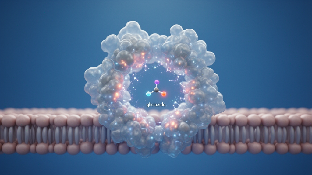
Pathogenic variants in the ABCC8 gene can alter the structure and drug sensitivity of sulfonylurea receptor 1, but a new computational study suggests that the diabetes medicine gliclazide can still bind to most of the 18 variants examined. Researchers combined molecular docking, 50-nanosecond molecular dynamics simulations and molecular mechanics–Poisson–Boltzmann surface area calculations to assess how the mutations affect the receptor’s conformational behavior and interaction with gliclazide. Across the models, the drug retained a broadly conserved binding pose, and none of the variants abolished its predicted association with the receptor. Phe591Leu produced the most stable simulation profile, whereas Asp1471Asn showed the greatest structural deviation. The authors caution that these findings are computational and require validation in functional channel assays and clinical studies.
ABCC8 encodes SUR1, the regulatory subunit of the ATP-sensitive potassium channel, or KATP, in pancreatic beta cells. The channel helps control insulin secretion by responding to cellular energy and nucleotide signals. Loss-of-function variants can impair channel opening and cause persistent hyperinsulinemic hypoglycemia, whereas gain-of-function variants can limit insulin release and contribute to diabetes or neonatal diabetes. SUR1 is also the principal pharmacological target of sulfonylureas. Gliclazide binds to this subunit and promotes channel closure, depolarizing the beta-cell membrane, opening voltage-gated calcium channels and triggering insulin release. Because ABCC8 variants occur in several diabetes-related disorders, their consequences for gliclazide binding may be relevant to drug selection and response.
The researchers assembled a dataset of 18 missense variants associated with type 2 diabetes from ClinVar Miner and ClinVar, retaining variants with at least two review stars and multiple submitters. The set included substitutions distributed throughout SUR1, including the gain-of-function Arg1182Trp and numerous loss-of-function variants linked to hyperinsulinism, neonatal diabetes or type 2 diabetes. They obtained the human wild-type sequence from UniProt and the corresponding structural model from the AlphaFold Protein Structure Database, then used AlphaFold 3 to model each mutant. Quality assessment with QMEANDisCo and MolProbity indicated that the models were suitable for downstream analysis: mutant global scores were close to the wild-type value, and more than 95% of residues in every model occupied favored Ramachandran regions. The variant models nevertheless had higher clash scores than the wild-type model, revealing localized differences in atomic contacts that could influence ligand interactions.
Molecular docking was performed with HADDOCK, using a predicted binding pocket to guide placement of gliclazide, diazoxide and metformin. Metformin served as a negative control because it lacks the sulfonylurea pharmacophore, while diazoxide provided a reference for another SUR1 ligand with opposite functional effects on channel gating. Gliclazide ranked as the strongest binder throughout the wild-type and mutant models, with predicted binding free energies of roughly −7 to −8 kilocalories per mole. The strongest docking result was for Phe591Leu at −7.95 kcal/mol, while Arg526Cys, Asp1471Asn, Arg74Gln and Glu128Lys also yielded favorable complexes. Most docked poses shared an Arg1144 hydrogen bond with gliclazide, while the wild type also interacted with Tyr1293. Mutants differed in aromatic and sulfur-mediated contacts involving Trp1296, Phe591, Tyr1293 or His584, showing how residue substitution could alter the local interaction network without eliminating ligand recognition.
The researchers advanced selected complexes into 50-nanosecond all-atom molecular dynamics simulations under explicit-solvent, physiological ionic conditions. Root-mean-square deviation, a measure of structural displacement, generally rose during the first 10 nanoseconds and then approached a plateau, suggesting equilibration rather than continuous destabilization. The wild-type gliclazide complex had a mean RMSD of 0.64 ± 0.13 nanometers. Phe591Leu was more stable still at 0.32 ± 0.04 nm, and Arg74Gln and Arg526Cys also produced relatively consistent trajectories. Asp1471Asn was the clearest outlier, with a mean RMSD of 0.92 ± 0.30 nm and a late rise to approximately 1.6–1.8 nm near the end of the run. That pattern indicated the greatest departure from the initial bound conformation, even though the ligand remained spatially associated with the receptor.
Other trajectory measures supported the general stability of the protein–ligand complexes. Mean residue fluctuations ranged from 0.17 to 0.27 nanometers, and the largest peaks occurred mainly in flexible loops and terminal regions outside the defined ligand-binding pocket. The radius of gyration stabilized after an early reduction, indicating that the global fold remained compact without sustained expansion. The wild-type gliclazide complex had a mean radius of gyration of 4.30 ± 0.02 nanometers, while Phe591Leu was slightly more compact at 4.21 ± 0.02 nanometers. Solvent-accessible surface area also declined during equilibration and then leveled off, with Phe591Leu showing the lowest mean value at 721.79 ± 20.49 square nanometers. These findings suggest that mutations in the tested set did not broadly destabilize the receptor’s fold or dislodge gliclazide over the simulation interval.
Ligand positioning and hydrogen bonding further distinguished the complexes. The mean distance between the ligand and receptor centers of mass remained within a relatively narrow range for most gliclazide complexes, indicating persistent association. Phe591Leu showed the least variation in this distance, while Asp1471Asn had the smallest average separation but the least stable global structure, showing that close proximity alone does not guarantee an intact complex. Phe591Leu also formed the most persistent polar contacts, averaging 2.67 hydrogen bonds and reaching a maximum of six. The wild-type complex averaged only 0.51 hydrogen bonds, and Arg526Cys averaged 0.19. These observations suggest that the Phe591Leu substitution may reinforce the bound state through a combination of compact protein geometry, consistent ligand placement and stronger hydrogen-bonding, even though the exact contribution of each interaction requires experimental testing.
Binding free-energy calculations gave a more nuanced picture than the structural stability metrics. MM-PBSA estimated the most favorable energy for wild-type gliclazide at −21.86 ± 2.70 kcal/mol, compared with −10.00 ± 3.20 for wild-type diazoxide and −9.34 ± 2.17 for wild-type metformin. Among the mutants, Arg74Gln had the most favorable gliclazide estimate at −19.46 ± 3.33 kcal/mol, followed by Phe591Leu at −18.95 ± 4.93 and Asp1471Asn at −18.18 ± 3.09. Arg526Cys and Glu128Lys were less favorable at −16.86 ± 3.81 and −16.34 ± 2.46 kcal/mol. Thus, every tested variant retained predicted binding but showed a less favorable free energy than the wild type. Phe591Leu illustrates why stability and affinity are not interchangeable: it had the most stable dynamics and strongest hydrogen bonding, yet its estimated binding energy remained slightly weaker than that of the wild type.
The biological interpretation remains tentative because docking and molecular dynamics describe interactions with a folded receptor and do not reproduce the full life cycle of a human KATP channel. SUR1 channels are assembled with Kir6.2 core subunits, and the simulations assumed homomeric mutant assemblies rather than the heteromeric arrangements found in many patients with compound-heterozygous mutations. They also cannot directly model whether a mutant receptor reaches the cell surface. That limitation is particularly relevant to Asp1471Asn, which has been associated with congenital hyperinsulinism and may impair channel folding or trafficking. A receptor retained in the endoplasmic reticulum could be unavailable to gliclazide even if its isolated protein model binds the drug. Future work should therefore express wild-type and mutant SUR1/KATP channels in cells, quantify sulfonylurea sensitivity with electrophysiology and derive concentration–response relationships, while also examining clinical outcomes in affected individuals.
Overall, the study portrays gliclazide as a robust computational binder of the pathogenic ABCC8 variants tested, but not as a variant-independent drug in every biological setting. The preserved Arg1144 anchor and recurring contacts in the sulfonylurea pocket help explain why the ligand remained associated with most models, while Phe591Leu emerged as a particularly stable complex and Asp1471Asn as the most likely to disturb structural behavior. The central message is that ABCC8 single-nucleotide variants can fine-tune SUR1 dynamics and modify predicted gliclazide affinity without eliminating binding. Establishing whether those differences translate into altered insulin secretion, channel closure or treatment response will require direct functional measurements and clinical validation rather than reliance on molecular simulation alone.
Subject of Research: Molecular simulation of how pathogenic ABCC8 variants affect structural dynamics and gliclazide binding to SUR1
Article Title: Structural dynamics of pathogenic ABCC8 single-nucleotide variants and their interactions with gliclazide
Article References: Simatupang, S. T., Dermawan, D., Nadia, Tan, S., Yulandi, A., & Tjandrawinata, R. R. (2026). Structural dynamics of pathogenic ABCC8 single-nucleotide variants and their interactions with gliclazide. Pharmacia, 73, Article e185272. https://doi.org/10.3897/pharmacia.73.e185272
Image Credits: AI Generated
DOI: 10.3897/pharmacia.73.e185272
Keywords: ABCC8, SUR1, gliclazide, type 2 diabetes, KATP channel, molecular dynamics, molecular docking, MM-PBSA, drug response, sulfonylurea receptor, insulin secretion, genetic variants
Cite Scienmag News
Juliet Wilcox. (October 20, 2026). Pathogenic ABCC8 variants mostly preserve gliclazide binding, simulations show. Scienmag. https://scienmag.com/pathogenic-abcc8-variants-mostly-preserve-gliclazide-binding-simulations-show/
Juliet Wilcox. "Pathogenic ABCC8 variants mostly preserve gliclazide binding, simulations show." Scienmag, 20 October 2026, https://scienmag.com/pathogenic-abcc8-variants-mostly-preserve-gliclazide-binding-simulations-show/. Accessed 20 October 2026.
Juliet Wilcox. "Pathogenic ABCC8 variants mostly preserve gliclazide binding, simulations show." Scienmag. October 20, 2026. https://scienmag.com/pathogenic-abcc8-variants-mostly-preserve-gliclazide-binding-simulations-show/


