


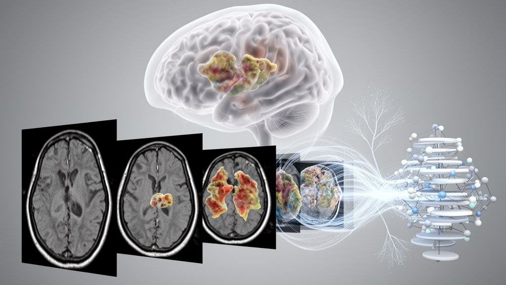
A unified artificial-intelligence system developed for brain-tumor MRI analysis achieved classification accuracies above 98% and Dice segmentation scores above 81% on two public datasets, according to a study published in Frontiers in Neuroscience. The model, called UAC-Net, performs two related tasks in a single framework: identifying whether a scan contains a glioma, meningioma or pituitary tumor, and producing a pixel-level map of the tumor. On the Brain Tumor dataset, it reached 98.53% accuracy and an 81.39% Dice coefficient. On the larger BRISC 2025 dataset, its accuracy rose to 99.07%, while Dice reached 86.61%. These are image-analysis results from publicly available datasets, not evidence that the system is ready for independent clinical use.
The design addresses a common inefficiency in medical-image analysis, where tumor classification and segmentation are often performed as separate tasks. Classification requires image-level information that distinguishes tumor categories, whereas segmentation requires detailed spatial information about lesion boundaries. UAC-Net uses a shared U-Net-style encoder so that both branches can learn from a common representation, while separate heads retain the different outputs required by the tasks. The segmentation branch reconstructs a tumor mask through progressive upsampling; the classification branch converts the deepest enhanced feature map into a global semantic vector through global average pooling and then predicts probabilities for the three tumor classes. The authors argue that pixel-level structure and image-level tumor semantics can be complementary during joint optimization.
Two specialized modules form the network’s principal technical contribution. The Aggregated Contextual Attention module, or ACA, is designed to combine local information with wider contextual cues. Its local sliding-window attention examines nearby pixels, helping preserve fine lesion structure, while a spatially downsampled global-attention path captures longer-range relationships. Learnable query embeddings give the attention calculation additional flexibility, a positional-attention matrix introduces a dynamic spatial bias, and a learnable scaling factor stabilizes the length-scaled cosine calculation when feature-map dimensions differ. The authors describe this dual-path arrangement as a way to emphasize tumor-related regions without allowing global context to obliterate local boundary information.
The second module, Adjacent Layer Multi-scale Feature Fusion, or ALMFF, addresses the semantic mismatch that can arise when features from different depths of an encoder are combined. Shallower maps contain more spatial and edge detail, while deeper maps carry stronger semantic information but have reduced resolution. ALMFF first adjusts the deeper adjacent feature with transposed convolution, then processes the aligned features through parallel 3-by-3 and 5-by-5 convolution branches. The smaller kernels capture local detail, whereas the larger kernels cover broader context. Element-wise multiplication combines their responses, and a residual connection preserves information and stabilizes propagation. In the complete network, ACA enhances these fused features before they enter either the segmentation decoder or the classification pathway.
The experiments used two publicly available collections of T1-weighted contrast-enhanced MRI scans. The Brain Tumor dataset contained 3,064 images with corresponding masks: 1,426 glioma images, 708 meningioma images and 930 pituitary-tumor images. The BRISC 2025 segmentation subset contained 4,793 images and 4,793 binary masks, comprising 1,401 glioma images, 1,635 meningioma images and 1,757 pituitary-tumor images. Images were resized to 224 by 224 pixels, replicated from one to three input channels and normalized. Random horizontal and vertical flipping and random intensity shifts were applied online during training only. Each dataset was divided into mutually exclusive training and testing sets at an 8:2 ratio, producing 2,450 training and 614 test images for Brain Tumor and 3,933 training and 860 test images for BRISC 2025.
Training combined a classification cross-entropy loss with a segmentation objective formed from binary cross-entropy and Dice loss. The total loss weighted segmentation and classification at initial values of 2 and 1, respectively, with the weights automatically updated from validation performance during training. The researchers used the Adam optimizer with an initial learning rate of 0.0001, a batch size of 8 and a maximum of 160 epochs, alongside a learning-rate scheduler and early stopping. All experiments ran on one workstation with 32 GB of random-access memory and an NVIDIA RTX 4060 Ti graphics processor with 16 GB of memory. UAC-Net contained 74.97 million parameters and was reported as requiring 36.52 gigaflops. Reported training times were 1.34 hours for Brain Tumor and 2.14 hours for BRISC 2025, while testing took 19 and 30 seconds, respectively.
Against four comparison architectures evaluated with the same splits and training conditions, UAC-Net produced the strongest overall results on both datasets. On Brain Tumor, conventional U-Net achieved 97.39% classification accuracy and a 78.56% Dice coefficient, Attention U-Net achieved 98.53% and 79.55%, SwinUNet reached 94.14% and 70.57%, and TransUNet reached 96.42% and 76.33%. UAC-Net’s Dice of 81.39% and intersection-over-union score of 72.66% exceeded those baselines, while its 95% Hausdorff distance of 5.782 millimetres was lower than the competing values. The margin was narrower on BRISC 2025: U-Net obtained a Dice score of 85.91%, compared with 86.61% for UAC-Net, but UAC-Net also had a lower boundary error of 4.713 millimetres and an IoU of 79.63%.
Ablation experiments on the Brain Tumor dataset showed that the two modules did not contribute equally in isolation. Adding ACA to the baseline raised accuracy from 97.39% to 98.21%, increased Dice from 78.56% to 80.55% and reduced HD95 from 7.731 to 6.141 millimetres. Adding ALMFF alone increased accuracy only to 97.56% and reduced Dice to 76.45%, with HD95 worsening to 9.478 millimetres. Combining both produced the best result: 98.53% accuracy, 81.39% Dice, 72.66% IoU and an HD95 of 5.782 millimetres. The authors interpret this pattern as evidence that adjacent-layer fusion can introduce redundant or conflicting responses unless contextual attention guides it. ALMFF performed best when paired with ACA, which they report as the dominant module in the ablation.
Further ablations distinguished UAC-Net from other attention and fusion choices. Squeeze-and-Excitation recalibration improved baseline Dice to 79.53%, while CBAM and a Transformer replacement reduced it to 75.72% and 75.56%, respectively. With ACA held constant, ALMFF outperformed simple concatenation, attention gates and atrous spatial pyramid pooling, achieving 81.39% Dice and the lowest HD95 of 5.782 millimetres. A separate comparison of task structure also indicated complementary multi-task learning: a classification-only version reached 98.20% accuracy, a segmentation-only version reached 72.93% Dice, and the joint model reached 98.53% accuracy and 81.39% Dice. Five repeated runs on Brain Tumor produced a mean accuracy of 98.42% plus or minus 0.22% and mean Dice of 81.50% plus or minus 0.57%, with all reported variation remaining modest. Grad-CAM++ visualizations showed high-activation regions concentrated mainly over tumor-related areas, although such images provide qualitative rather than independent clinical validation.
Several limitations temper the results. Both datasets contain only three tumor classes and use a single MRI contrast, so the study does not establish performance across the wider spectrum of brain tumors or clinical sequences. Very small lesions, weak lesion-to-background contrast and irregular boundaries remain difficult, and the authors note that repeated downsampling can weaken fine spatial information. The two public datasets were divided internally into training and testing sets, rather than undergoing prospective, multicenter or external clinical evaluation. Scanner differences, acquisition protocols, patient populations and annotation variability could therefore affect performance in practice. The model also remains substantial at nearly 75 million parameters. The findings support UAC-Net as a promising technical approach to joint MRI tumor typing and localization, but its high benchmark scores do not demonstrate clinical benefit, diagnostic safety or readiness for deployment without further validation.
Subject of Research: Multi-task deep learning for brain-tumor MRI classification and segmentation
Article Title: UAC-Net: a unified multi-task network with aggregated contextual attention and cross-layer fusion for brain tumor segmentation and classification
Article References: Cai, C., Zhang, J., Zhang, D., Chen, W., Tan, X., Du, G., & Jiang, H. (2026). UAC-Net: a unified multi-task network with aggregated contextual attention and cross-layer fusion for brain tumor segmentation and classification. Frontiers in Neuroscience, 20, Article 1986322. https://doi.org/10.3389/fnins.2026.1986322
Image Credits: AI Generated
DOI: 10.3389/fnins.2026.1986322
Keywords: brain tumors, MRI analysis, deep learning, multi-task learning, image segmentation, tumor classification, contextual attention, feature fusion, U-Net, UAC-Net, unified, multi-task
Cite Scienmag News
Cassandra Pierce. (October 20, 2026). UAC-Net links brain-tumor typing and MRI segmentation in one model. Scienmag. https://scienmag.com/uac-net-links-brain-tumor-typing-and-mri-segmentation-in-one-model/
Cassandra Pierce. "UAC-Net links brain-tumor typing and MRI segmentation in one model." Scienmag, 20 October 2026, https://scienmag.com/uac-net-links-brain-tumor-typing-and-mri-segmentation-in-one-model/. Accessed 20 October 2026.
Cassandra Pierce. "UAC-Net links brain-tumor typing and MRI segmentation in one model." Scienmag. October 20, 2026. https://scienmag.com/uac-net-links-brain-tumor-typing-and-mri-segmentation-in-one-model/


