


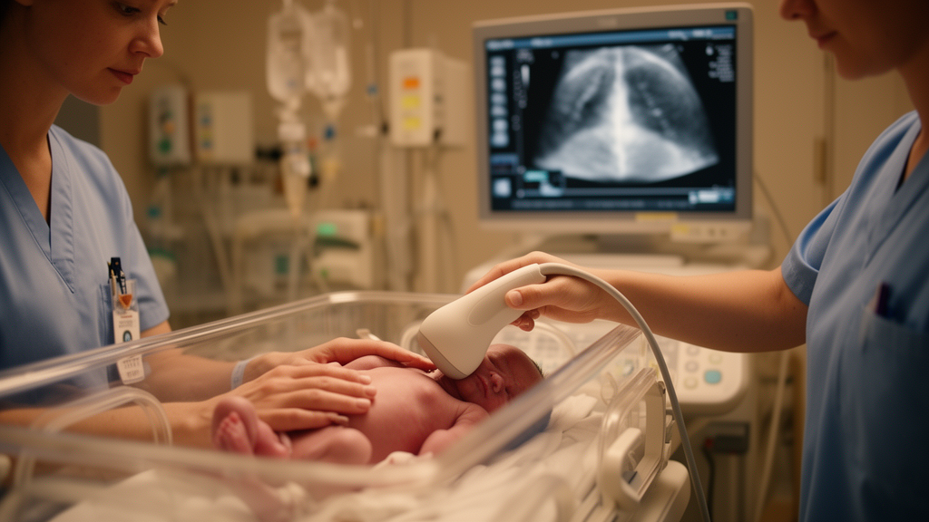
In the neonatal intensive care unit, few tools are as quietly transformative as a handheld ultrasound probe. A new narrative review published in the Journal of Perinatology by Nicole Stephenson, Cara Beth Carr, Daniel Park, and Matthew Laughon of the University of North Carolina School of Medicine synthesizes the rapidly expanding evidence base for lung ultrasound in preterm infants, arguing that the bedside technique is poised to reshape how clinicians diagnose respiratory distress syndrome, decide when to give surfactant, judge extubation readiness, and predict which babies will go on to develop bronchopulmonary dysplasia. Because the method relies on sound waves rather than ionizing radiation, it can be repeated as often as needed, a decisive advantage in a population whose fragile lungs may require imaging dozens of times across a hospital stay that can stretch for months.
The physics behind lung ultrasound is deceptively simple. The probe, placed on the chest wall, cannot image aerated lung directly, because air scatters ultrasound almost completely. Instead, clinicians read artifacts: the shimmering, to-and-fro movement of the pleural line known as lung sliding, the horizontal reverberation lines called A-lines that indicate normal aeration, and the vertical comet-tail artifacts known as B-lines that signal fluid or collapsed, non-aerated tissue beneath the probe. In preterm infants with respiratory distress syndrome, a disease of surfactant deficiency in which alveoli collapse, the typical pattern includes diffuse B-lines, thickened or irregular pleural lines, and consolidated, tissue-like areas where lung has lost its air entirely. As surfactant therapy recruits alveoli and aeration returns, these findings evolve in real time, giving clinicians a functional, dynamic picture of the lung rather than the static snapshot offered by a chest radiograph.
That dynamism is precisely what makes the technology attractive for one of the most consequential decisions in neonatology: whether and when to administer surfactant. The review highlights a multicenter pragmatic study by Raimondi and colleagues, published in Chest in 2021, in which lung ultrasound findings were used to guide surfactant administration in preterm infants with respiratory distress. Related work by De Martino and colleagues showed that a lung ultrasound score obtained shortly after birth could predict the need for surfactant in extremely preterm neonates, and Badurdeen and colleagues demonstrated that ultrasound performed during newborn resuscitation predicted surfactant requirement in very and extremely preterm infants. Even more striking, the ULTRASURF randomized controlled trial by Rodriguez-Fanjul and colleagues tested early surfactant replacement guided by lung ultrasound against conventional management in preterm newborns with respiratory distress syndrome, providing some of the strongest evidence that the modality can drive treatment decisions rather than merely describe disease.
The scoring systems at the heart of these studies deserve technical attention. Most divide the chest into a grid of zones, commonly four, six, eight, or twelve, and assign each zone a numerical grade based on the worst finding present: zero for normal aeration with A-lines, rising through increasing numbers of well-separated B-lines, to coalescent B-lines, and finally to large consolidations with air bronchograms for the most severely affected regions. Summing the zone scores yields a global aeration index. The review compares these protocols explicitly, noting that the choice of zone count affects sensitivity, examination time, and reproducibility. A 2025 systematic review by Sartorius, Brunet, and De Luca catalogued the characteristics of the many neonatal lung ultrasound scores now in circulation, and a 2022 narrative review by Corsini and colleagues mapped how they are used in clinical practice. The proliferation of scoring schemes, the authors caution, is both a sign of the field’s vitality and a source of its fragmentation.
Perhaps the most clinically consequential application is the prediction of bronchopulmonary dysplasia, the chronic lung disease of prematurity that remains one of the most common and feared outcomes for infants born extremely early. Because BPD is diagnosed clinically at 36 weeks postmenstrual age, clinicians often spend weeks caring for an infant without a clear sense of that infant’s long-term pulmonary trajectory. Lung ultrasound offers a potential early warning system. A prospective multicenter diagnostic accuracy study by Alonso-Ojembarrena and colleagues, published in Chest in 2021, evaluated the predictive value of lung ultrasound scores for developing BPD, and a multicenter longitudinal cohort study by Loi and colleagues in the American Journal of Respiratory and Critical Care Medicine showed that serial ultrasound could monitor extremely preterm infants and predict BPD. Oulego-Erroz and colleagues demonstrated that early assessment of lung aeration using an ultrasound score could serve as a biomarker of subsequent BPD, and Mohamed and colleagues reported similar predictive performance in a prospective diagnostic cohort of extreme preterm neonates.
The prognostic reach of the technique extends beyond the diagnosis itself. Zong and colleagues showed that a lung ultrasound score could predict the severity of bronchopulmonary dysplasia in neonates born at or before 25 weeks of gestation, while Aldecoa-Bilbao and colleagues characterized distinct ultrasound patterns and predictors in very preterm infants with the disease. In a comparison that underscores the score’s discriminative power, Alonso-Ojembarrena and colleagues found that lung ultrasound outperformed NT-proBNP, a widely used biomarker, in predicting moderate to severe BPD. Modified scores have been developed specifically for the BPD population, and Hoshino and colleagues used lung ultrasound to predict respiratory outcomes in patients with established disease. Savoia and colleagues even showed that the ultrasound score follows the trajectory of chronic pulmonary insufficiency of prematurity into early infancy, suggesting the technique can track disease evolution well beyond the NICU.
That longer-term horizon is where the review points toward the future. Shen and colleagues reported that a modified lung ultrasound score for BPD predicted late respiratory outcomes in preterm infants, and a 2024 study published in JAMA Network Open by Bonadies and colleagues linked lung ultrasonography scores in preterm infants to respiratory outcomes at age two. Stephenson and her own group had previously published work on outpatient assessment of bronchopulmonary dysplasia using point-of-care lung ultrasound, extending the modality from the intensive care unit into the clinic. The review also notes emerging data on monitoring pulmonary edema: Kasniya and colleagues assessed pulmonary edema in neonates with chronic lung disease before and after diuretic therapy, and a pilot study by Alonso-Ojembarrena and colleagues examined lung ultrasound scores and diuretics in infants born before 32 weeks. Together, these studies sketch a vision of lung ultrasound as a continuous, longitudinal readout of pulmonary health from the delivery room through early childhood.
The modality’s diagnostic repertoire is not limited to surfactant decisions and BPD prediction. The review situates lung ultrasound within the broader landscape of neonatal pulmonary imaging, citing evidence that it can diagnose and differentiate transient tachypnea of the newborn from respiratory distress syndrome, detect pneumothorax with performance that a systematic review by Fei and colleagues suggested may make it a better first choice than radiography, and identify pneumonia, including in neonates with fungal infections. It can also inform weaning from mechanical ventilation: Soliman and colleagues used lung ultrasound to predict extubation readiness in preterm infants, and El Amrousy and colleagues evaluated its value in predicting weaning success in ventilated neonates. Because extubation failure carries real costs in a preterm population, a bedside tool that quantifies aeration and diaphragm-adjacent lung recruitment at the moment of decision could meaningfully change practice.
Yet the review is candid about the field’s limitations. Interpretation of the existing evidence is constrained by heterogeneity in acquisition protocols, scoring systems, and the timing of assessments, and by the fact that lung ultrasound is fundamentally operator dependent. A score obtained with a twelve-zone protocol by an experienced sonographer is not interchangeable with a four-zone scan performed by a trainee, and the timing of an assessment, whether in the first minutes of life, during the first days of respiratory distress, or in the weeks before a BPD diagnosis, can dramatically alter its predictive meaning. Most studies to date have been single center or modest in size, and few have tested whether ultrasound-guided management actually improves hard outcomes such as mortality, ventilator days, or long-term neurodevelopment, as opposed to simply changing when surfactant is given. The authors call explicitly for further multicenter studies with standardized protocols to define the role of lung ultrasound in routine neonatal respiratory care.
Even with those caveats, the trajectory is unmistakable. A technique that requires no radiation, sits at the bedside, delivers results in minutes, and can be repeated without limit addresses nearly every weakness of the chest radiograph in neonatology, where transport to the X-ray suite is hazardous, images are snapshots rather than movies, and cumulative dose is a genuine concern for infants who may need hundreds of days of intensive care. If ongoing trials confirm that standardized lung ultrasound scores can reliably triage surfactant, time extubation, and stratify BPD risk early enough to change management, the handheld probe may become as fundamental to the preterm infant’s care as the stethoscope once was, turning the NICU’s most vulnerable lungs into an open book read in real time.
Subject of Research: Lung ultrasound applications for respiratory distress syndrome and bronchopulmonary dysplasia in preterm infants
Article Title: Applications of lung ultrasound in preterm infants: implications for respiratory distress syndrome and bronchopulmonary dysplasia
Article References: Stephenson, N., Carr, C. B., Park, D., & Laughon, M. (2026). Applications of lung ultrasound in preterm infants: implications for respiratory distress syndrome and bronchopulmonary dysplasia. Journal of Perinatology. https://doi.org/10.1038/s41372-026-02849-w
Image Credits: AI Generated
DOI: 10.1038/s41372-026-02849-w
Keywords: lung ultrasound, preterm infants, respiratory distress syndrome, bronchopulmonary dysplasia, surfactant therapy, neonatal intensive care, medical imaging, lung ultrasound score, extubation readiness, predictive markers, neonatology, point-of-care ultrasound
Cite Scienmag News
Harold Sullivan. (October 19, 2026). Lung Ultrasound Emerges as a Radiation-Free Window Into Preterm Infant Lungs. Scienmag. https://scienmag.com/lung-ultrasound-emerges-as-a-radiation-free-window-into-preterm-infant-lungs/
Harold Sullivan. "Lung Ultrasound Emerges as a Radiation-Free Window Into Preterm Infant Lungs." Scienmag, 19 October 2026, https://scienmag.com/lung-ultrasound-emerges-as-a-radiation-free-window-into-preterm-infant-lungs/. Accessed 19 October 2026.
Harold Sullivan. "Lung Ultrasound Emerges as a Radiation-Free Window Into Preterm Infant Lungs." Scienmag. October 19, 2026. https://scienmag.com/lung-ultrasound-emerges-as-a-radiation-free-window-into-preterm-infant-lungs/


