


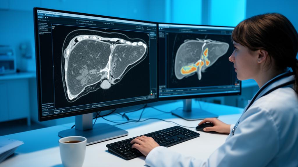
When colorectal cancer spreads to the liver and surgeons can no longer remove the tumors, patients and their doctors face an anxious guessing game: will months of chemotherapy actually hold the disease in check, or will the cancer keep growing despite treatment? A team of researchers in Guangzhou, China, has now built a machine learning tool that tries to answer that question before the first infusion begins, using nothing more than a routine contrast-enhanced CT scan and a handful of blood test results. The study, published in BMC Medical Imaging, describes a radiomics-based model that analyzes every measurable liver metastasis on a scan and predicts whether a patient will still be free of disease progression twelve months after starting first-line chemotherapy.
The approach, known as radiomics, rests on a simple but powerful idea: medical images contain far more information than the human eye can extract. Radiomics algorithms convert the pixel patterns inside a tumor into hundreds of quantitative features, capturing properties such as texture heterogeneity, gray-level distribution, and the statistical relationships between neighboring pixels. These features can reflect underlying biology, including how disorganized and aggressive a tumor’s cells are, long before the tumor visibly shrinks or grows. In this study, the researchers pushed the concept further by analyzing not just the largest lesion, as most prior radiomics studies have done, but every measurable liver metastasis in each patient, capturing the full spatial diversity of the disease.
The retrospective cohort included 159 patients with unresectable colorectal liver metastases who all received contrast-enhanced CT before beginning first-line chemotherapy. The team divided the patients into a training cohort of 127 individuals and an internal hold-out set of 32, using stratified sampling to preserve the balance of outcomes between the groups. For every patient, radiologists separately segmented all measurable liver metastases on both the arterial phase and the portal venous phase of the CT scan, the two contrast-enhanced windows that highlight different aspects of tumor blood supply. From each segmented lesion, the pipeline extracted radiomic features, which were then aggregated across lesions to produce a single multi-lesion signature per imaging phase per patient.
One of the study’s most distinctive technical contributions lies in how the researchers chose which features to keep. High-dimensional radiomics datasets typically contain far more variables than patients, creating a serious risk that models will memorize noise rather than learn genuine biological signals. Instead of relying solely on conventional statistical filters or the widely used LASSO regression, the team applied a method called CRITIC, short for Criteria Importance Through Intercriteria Correlation, which objectively weights each feature by combining its contrast across patients with the degree to which it overlaps informationally with other features. They then fused these weights into a multi-criteria decision making framework, producing a feature selection pipeline the authors call CRITIC_MCDM. In head-to-head comparisons against alternative selection methods, this pipeline achieved the highest numerical AUC, although the differences did not reach statistical significance.
With curated features in hand, the researchers trained multiple machine learning classifiers on three separate data streams: the arterial-phase radiomics, the portal venous-phase radiomics, and a set of clinical variables that included the tumor markers CA19-9 and CEA along with whether the primary colorectal tumor had been surgically resected. Rather than betting on any single algorithm, they integrated the base learners using a performance-weighted soft-voting ensemble, a strategy in which each model’s vote on a patient’s outcome is scaled by how well that model performed during training. The resulting trimodal model, abbreviated AVC for arterial, venous, and clinical, delivered the study’s headline result: in the internal hold-out set, it achieved an AUC of 0.792, with a 95 percent confidence interval stretching from 0.625 to 0.933.
An AUC of roughly 0.79 places the model in the territory of clinically useful but not yet definitive discrimination, meaning it can meaningfully separate patients likely to remain progression-free at twelve months from those likely to progress early, while still misclassifying a substantial minority. The authors are candid about this. Their statistical analysis showed the study was underpowered for formal comparisons between competing models, and the confidence interval around the headline AUC is wide enough to encompass performance ranging from modest to excellent. Decision curve analysis, a technique that quantifies the net clinical benefit of acting on a model’s predictions across a range of decision thresholds, suggested a favorable net benefit within part of the clinically relevant threshold range, but the researchers explicitly note that this finding requires confirmation in larger external cohorts.
Interpretability was a central design goal rather than an afterthought. Using Shapley additive explanations, or SHAP, a technique borrowed from cooperative game theory that assigns each feature a quantified contribution to every individual prediction, the team identified which signals drove the model’s outputs. Among the most influential were first-order statistics from the arterial phase, exemplified by a feature labeled A_FO_10Pct_sum, which summarizes the distribution of pixel intensities within lesions during the phase when arterial blood supply dominates. From the portal venous phase, first-order skewness and gray-level dependence matrix features, such as V_FO_Skew_mean, captured the asymmetry and structural coarseness of tumor texture. Alongside these image-derived signals, the blood biomarkers CA19-9 and CEA and the status of primary tumor resection carried substantial predictive weight, confirming that the model integrates imaging biology with established clinical prognostic factors rather than replacing them.
The multi-lesion design deserves particular attention because it addresses a well-known blind spot in oncology imaging. Colorectal liver metastases are rarely uniform; a single patient’s liver may harbor lesions with markedly different textures and enhancement patterns, reflecting genuine biological heterogeneity within the same tumor population. Analyzing only the largest lesion risks discarding this diversity, which may itself carry prognostic information. By segmenting and aggregating features across all measurable metastases, the model builds a portrait of the disease as a whole, an approach the authors argue better reflects the biology that determines how the cancer responds to systemic chemotherapy.
The practical appeal of the framework lies in its accessibility. Contrast-enhanced CT is already a standard part of staging and surveillance for colorectal cancer patients worldwide, and CA19-9 and CEA are routinely measured in clinical practice. A validated version of this model could therefore be deployed without new imaging protocols, new scans, or invasive biopsies, offering oncologists a quantitative risk estimate at the very start of treatment. Such a tool could help stratify patients in clinical trials, guide the intensity of surveillance, and support conversations about treatment expectations, all while remaining fully interpretable through SHAP-based explanations of each prediction.
Still, the path from a retrospective single-center study to bedside decision support is long. The hold-out set of 32 patients is small, the cohort comes from a single institution, and the authors themselves emphasize that prospective external validation is needed before any clinical application. The study also excluded treatment-related variables from a reduced version of the model, underscoring the importance of ensuring that predictions reflect tumor biology rather than artifacts of the treatment pipeline. If larger, multi-center, prospective studies confirm the performance suggested here, multi-lesion radiomics could become a routine lens through which oncologists view metastatic liver disease, turning every staging CT scan into a data-rich forecast of the road ahead.
Subject of Research: A multi-lesion CT radiomics machine learning model for predicting progression-free survival in unresectable colorectal liver metastases treated with chemotherapy
Article Title: A multi-lesion radiomics model for predicting progression-free survival in patients with unresectable colorectal liver metastases undergoing chemotherapy
Article References: Wu, Y., Li, L., Liu, X., Zhang, B., Chen, Z., Zhu, L., Tao, L., Ren, Y., Hong, H., Zhang, L., Liu, B., Ma, H., Wang, X., & He, C. (2026). A multi-lesion radiomics model for predicting progression-free survival in patients with unresectable colorectal liver metastases undergoing chemotherapy. BMC Medical Imaging. https://doi.org/10.1186/s12880-026-02768-4
Image Credits: AI Generated
DOI: 10.1186/s12880-026-02768-4
Keywords: colorectal liver metastases, radiomics, machine learning, contrast-enhanced CT, progression-free survival, chemotherapy, feature selection, SHAP, ensemble learning, tumor heterogeneity, predictive medicine, cancer imaging
Cite Scienmag News
Nathaniel Bowman. (October 18, 2026). AI Reads Liver Scans to Predict Which Colorectal Cancer Patients Will Respond to Chemotherapy. Scienmag. https://scienmag.com/ai-reads-liver-scans-to-predict-which-colorectal-cancer-patients-will-respond-to-chemotherapy/
Nathaniel Bowman. "AI Reads Liver Scans to Predict Which Colorectal Cancer Patients Will Respond to Chemotherapy." Scienmag, 18 October 2026, https://scienmag.com/ai-reads-liver-scans-to-predict-which-colorectal-cancer-patients-will-respond-to-chemotherapy/. Accessed 18 October 2026.
Nathaniel Bowman. "AI Reads Liver Scans to Predict Which Colorectal Cancer Patients Will Respond to Chemotherapy." Scienmag. October 18, 2026. https://scienmag.com/ai-reads-liver-scans-to-predict-which-colorectal-cancer-patients-will-respond-to-chemotherapy/


