


A previously healthy 12-year-old girl walked into a clinic with a two-month history of persistent left-sided headache, progressive swelling around her left orbit, and a visibly protruding left eye. Over the following weeks, she was variously diagnosed with typhoid fever, then migraine, before a left cervical mass finally emerged. By the time clinicians pieced together the full picture, the true culprit was not an infection at all: it was a high-grade diffuse B-cell non-Hodgkin lymphoma that had been masquerading, clinically and radiologically, as a deep neck space infection. The case, published in BMC Pediatrics by a team from the University of Aleppo in Syria, is a striking reminder of how closely pediatric head and neck malignancies can imitate common infectious diseases—and how dangerous that imitation can be when it delays the correct diagnosis.
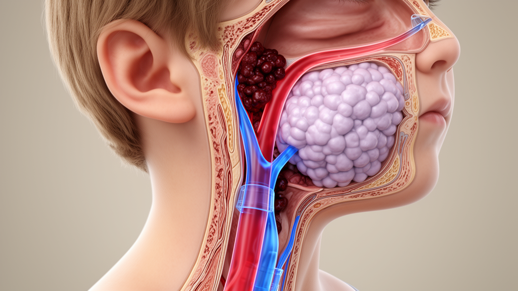
The clinical course deteriorated rapidly after a tonsillectomy and biopsy of a parapharyngeal mass. The girl developed acute respiratory distress, severe dysphagia, dysarthria, and an isolated lower motor neuron palsy of the hypoglossal nerve, the twelfth cranial nerve that controls tongue movement. These cranial neuropathies, combined with a deep neck mass, pushed the diagnostic team toward a presumed deep neck space infection. Contrast-enhanced computed tomography then revealed something more alarming: extensive thrombosis of the left sigmoid sinus and the left internal jugular vein, alongside the deep neck mass. The combination of an infectious-looking neck process and widespread venous clotting seemed internally consistent—an aggressive infection seeding the venous drainage of the brain and neck.
That internal consistency, the authors argue, is precisely what made the case such a diagnostic dilemma. Deep neck space infections are among the most common serious head and neck emergencies in children, and they can indeed be complicated by septic thrombophlebitis of the internal jugular vein, a condition historically known as Lemierre syndrome when it involves anaerobic bacteria. Cerebral venous sinus thrombosis, meanwhile, is a recognized complication of both infection and inflammatory disease in children. The presence of a cervical mass, cranial nerve dysfunction, and venous thrombosis therefore pointed convincingly toward an infectious etiology, and the patient was initially managed accordingly, with anticoagulation therapy successfully initiated to manage the extensive clotting.
The histopathology added yet another layer of confusion. Initial microscopic evaluation of the biopsy material was misinterpreted as histoplasmosis, a fungal infection that can involve the head and neck and can produce tissue reactions that overlap with lymphoid malignancies. It took expert slide re-evaluation and immunohistochemical characterization to definitively establish the correct diagnosis. The tumor cells showed diffuse positivity for CD20, a B-cell surface marker, and a Ki-67 proliferative index of approximately 90 percent—a figure indicating that nearly all of the tumor cells were actively dividing, consistent with a high-grade, rapidly proliferating B-cell lymphoma. Such a high proliferative fraction is characteristic of aggressive lymphomas like Burkitt lymphoma or diffuse large B-cell lymphoma, which are among the most common pediatric non-Hodgkin lymphomas.
The coexistence of lymphoma with extensive venous thromboembolism—specifically cerebral venous sinus thrombosis and internal jugular vein thrombosis—is uncommon and introduces profound diagnostic challenges, the authors note. Venous thrombosis in this case arose from two converging mechanisms: tumor-associated hypercoagulability, in which malignant cells release procoagulant factors and inflammatory cytokines that shift the blood toward clotting, and direct mechanical compression of the internal jugular vein and adjacent venous structures by the growing mass. This dual mechanism is well recognized in oncology but is rarely considered at the front end of a pediatric neck evaluation, where infection remains the default explanation for a cervical mass with cranial neuropathies.
The case highlights several technical lessons for clinicians evaluating children with head and neck pathology. Persistent headache in a child, particularly when progressive and unilateral, should never be dismissed as migraine without careful consideration of structural causes. Orbital swelling and proptosis, meanwhile, indicate pathology behind or around the eye that demands urgent imaging. Cranial neuropathies—an isolated hypoglossal palsy in this case—suggest involvement of the skull base or deep neck spaces where cranial nerves traverse, a territory that can be occupied by infection, inflammation, or malignancy. And cerebral venous sinus thrombosis in a child, while it can complicate infection, should also prompt consideration of underlying hypercoagulable states, including malignancy.
The discordance between clinical, radiological, and pathological findings is the central theme of the report. The clinical picture suggested infection. The radiology showed a deep neck mass with venous thrombosis, which could fit either infection or tumor. And the initial histopathology suggested a fungal organism. Each individual finding was internally plausible, but collectively they were contradictory—and it was only through early multidisciplinary reassessment and expert pathological review that the discordance was resolved. The authors emphasize that when such discordance exists, particularly in a pediatric patient, underlying malignancy must be actively considered rather than assumed away by the more familiar infectious explanations.
The diagnostic delay in this case—two months of alternative diagnoses including typhoid fever and migraine before the correct diagnosis was established—illustrates the cost of anchoring on common explanations. High-grade B-cell lymphomas in children are among the most rapidly proliferating human malignancies, and delays of even weeks can affect staging and treatment intensity. The patient was ultimately referred for definitive oncological care after anticoagulation was successfully managed, but the case underscores how easily a lymphoma can be mistaken for a deep neck infection, a histoplasmosis, or a simple vascular complication of an infectious process.
For the broader scientific community, the case adds to a growing literature on the ability of pediatric lymphomas to closely mimic deep neck infections clinically, radiologically, and histopathologically. The authors conclude that persistent headache, cranial neuropathies, cervical masses, or cerebral venous thrombosis in children should prompt consideration of underlying malignancy, particularly when clinical, radiological, and pathological findings are discordant. Early multidisciplinary reassessment and expert pathological review, they argue, are essential to avoid diagnostic delay and ensure timely oncological management—a lesson that could prove decisive for other children whose lymphomas are hiding behind the familiar face of an infection.
Subject of Research: Pediatric high-grade diffuse B-cell non-Hodgkin lymphoma mimicking a deep neck space infection with internal jugular vein and sigmoid sinus thrombosis: a diagnostic dilemma
Article Title: Pediatric high-grade diffuse B-cell non-Hodgkin lymphoma mimicking a deep neck space infection with internal jugular vein and sigmoid sinus thrombosis: a diagnostic dilemma
Article References: Alhussein, H., Eid, H., Al-ogaili, A., Fattouh, S., & Alabd, N. (2026). Pediatric high-grade diffuse B-cell non-Hodgkin lymphoma mimicking a deep neck space infection with internal jugular vein and sigmoid sinus thrombosis: a diagnostic dilemma. BMC Pediatrics. https://doi.org/10.1186/s12887-026-07644-2
Image Credits: AI Generated
DOI: 10.1186/s12887-026-07644-2
Keywords: Pediatric, high-grade, diffuse, B-cell, non-Hodgkin, lymphoma, mimicking, deep, neck, space, infection, internal
Cite Scienmag News
Ophelia Keating. (October 18, 2026). Pediatric high-grade diffuse B-cell non-Hodgkin lymphoma mimicking a deep neck space infection with internal jugular vein and sigmoid sinus thrombosis: a diagnostic dilemma. Scienmag. https://scienmag.com/pediatric-high-grade-diffuse-b-cell-non-hodgkin-lymphoma-mimicking-a-deep-neck-space-infection-with-internal-jugular-vein-and-sigmoid-sinus-thrombosis-a-diagnostic-dilemma/
Ophelia Keating. "Pediatric high-grade diffuse B-cell non-Hodgkin lymphoma mimicking a deep neck space infection with internal jugular vein and sigmoid sinus thrombosis: a diagnostic dilemma." Scienmag, 18 October 2026, https://scienmag.com/pediatric-high-grade-diffuse-b-cell-non-hodgkin-lymphoma-mimicking-a-deep-neck-space-infection-with-internal-jugular-vein-and-sigmoid-sinus-thrombosis-a-diagnostic-dilemma/. Accessed 18 October 2026.
Ophelia Keating. "Pediatric high-grade diffuse B-cell non-Hodgkin lymphoma mimicking a deep neck space infection with internal jugular vein and sigmoid sinus thrombosis: a diagnostic dilemma." Scienmag. October 18, 2026. https://scienmag.com/pediatric-high-grade-diffuse-b-cell-non-hodgkin-lymphoma-mimicking-a-deep-neck-space-infection-with-internal-jugular-vein-and-sigmoid-sinus-thrombosis-a-diagnostic-dilemma/


