


For decades, phytosterols—the plant-derived sterols found in vegetable oils, nuts, seeds, and grains—have been regarded as dietary bystanders. Unlike cholesterol, roughly half of which is absorbed from the intestine, only about five percent of phytosterols ever enter the bloodstream. The intestinal epithelial transporter Niemann-Pick C1-like 1, or NPC1L1, brings a small fraction across the epithelial barrier, but the heterodimeric ATP-binding cassette transporters ABCG5 and ABCG8 promptly pump most of it back into the gut lumen. As a result, phytosterol levels in mammalian tissues remain vanishingly low, and when the efflux transporters fail, as in the human genetic disorder sitosterolemia, phytosterols pile up abnormally and contribute to tendon xanthomas, premature atherosclerosis, thrombocytopenia, and hemolytic anemia. Maintaining low phytosterol levels, the conventional thinking holds, is essential for animal health.
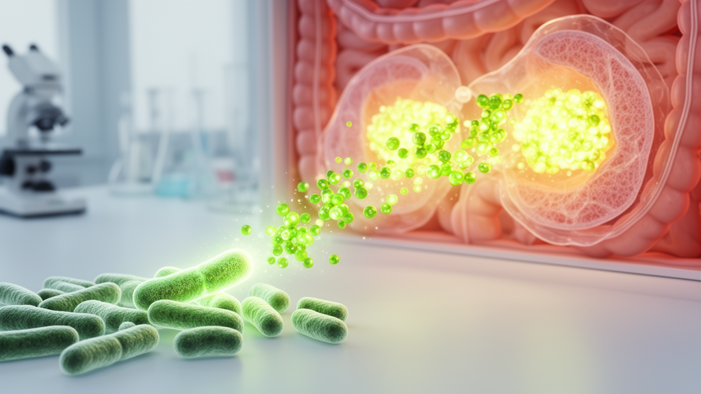
A new study published in iScience by Koki Tomita, Takeru Nakano, Satoshi Morozumi, Nobuyuki Okahashi, and Makoto Arita of Keio University and collaborating institutions now upends that picture. Through untargeted lipidomics data mining, the team identified phytosterol sulfates—sitosterol sulfate, campesterol sulfate, stigmasterol sulfate, and coprostanol sulfate—as metabolites produced by the gut microbiota rather than by the host. The researchers traced the responsible enzymes to bacterial sulfotransferase genes, designated Sults, carried by members of the Bacteroidota phylum. Crucially, they showed that these microbially generated sulfated sterols do not stay in the gut: they translocate into the circulation and accumulate in multiple mouse tissues in a microbiota- and age-dependent manner, revealing a previously unrecognized route by which gut microbes transform dietary sterols into metabolites that persist within the host.
The investigation began with a reanalysis of previously published untargeted fecal lipidome data from specific pathogen-free and germ-free mice. Alongside cholesterol sulfate, a well-known host-derived sterol sulfate, the mass spectrometry spectra revealed a series of molecules sharing a diagnostic sulfate ion fragment at mass-to-charge ratio 96.960. These were identified as sitosterol sulfate, campesterol sulfate, stigmasterol sulfate, and coprostanol sulfate. Cholesterol sulfate abundance was not significantly reduced in germ-free mice compared with specific pathogen-free mice, indicating only a minor microbial contribution to fecal cholesterol sulfate. In stark contrast, the phytosterol sulfates collapsed in the absence of microbes. Sitosterol sulfate and stigmasterol sulfate showed the strongest microbiota dependence, falling by 98.2 percent and 99.2 percent respectively in germ-free animals, while campesterol sulfate and coprostanol sulfate dropped by 87.4 percent and 64.6 percent. Reanalysis of fecal lipidomics from mice whose gut microbiota had been eliminated with an antibiotic cocktail confirmed the pattern, with sitosterol sulfate emerging as the most prominent microbiota-dependent phytosterol sulfate.
To pinpoint the producers, the team cultured ten representative Bacteroidota species anaerobically with cholesterol or sitosterol. Six species—Parabacteroides goldsteinii, Parabacteroides johnsonii, Bacteroides uniformis, Bacteroides fragilis, Bacteroides caccae, and Bacteroides thetaiotaomicron—produced both cholesterol sulfate and sitosterol sulfate, while four others, including Alistipes finegoldii and Porphyromonas gingivalis, produced neither. Homology searches using the amino acid sequence of the known sterol sulfotransferase from Parabacteroides merdae as a query revealed that all six producing bacteria harbored a homologous Sult gene, with sequence identities ranging from 75 to 98 percent. When each Sult homolog was heterologously expressed in Escherichia coli and incubated with sitosterol and the universal sulfate donor 3′-phosphoadenosine-5′-phosphosulfate, every enzyme generated sitosterol sulfate. Bacterial sitosterol sulfate production by Sults, the results demonstrate, is common among specific Bacteroidota species, several of which are major members of the human gut microbiota.
The study then probed whether the bacterial enzymes and their mammalian counterparts differ in substrate preference. The researchers compared the recombinant Bacteroides thetaiotaomicron Sult, designated BtSult, with mouse Sult2b1b and human SULT2B1b, the host enzymes that normally sulfate cholesterol. All three converted cholesterol to cholesterol sulfate, but their efficiency toward phytosterols diverged sharply. When cholesterol sulfate production was normalized to 100 percent, sitosterol sulfate production ratios were 51.6 percent for BtSult, 19.5 percent for mouse Sult2b1b, and just 11.4 percent for human SULT2B1b. BtSult also more efficiently converted campesterol, brassicasterol, and stigmasterol to their sulfated forms than either mammalian enzyme. Biochemical considerations help explain the difference: structural work on human SULT2B1b has suggested that the C-17 alkyl group of cholesterol directly contacts the Ile20 residue of the catalytic site, constraining substrate recognition, whereas bacterial Sults likely possess more room to accommodate the bulky alkyl side chains that distinguish phytosterols. Given the low efficiency of the mammalian enzymes and the modest abundance of phytosterol substrate reaching host tissues, the authors conclude that phytosterol sulfates present in host tissues are mostly derived from the gut microbiota.
The most striking finding concerns tissue distribution. Reanalyzing a large-scale untargeted lipidomic dataset spanning feces, plasma, and ten organs from specific pathogen-free and germ-free mice, the team detected sitosterol sulfate in nine tissues, including the liver, kidney, and plasma, in 19-month-old male mice. In all tissues, sitosterol sulfate intensity was approximately ten percent that of cholesterol sulfate, but the two molecules showed markedly different distribution patterns: cholesterol sulfate was present in the skin, mesentery, and skeletal muscle, where sitosterol sulfate was absent. Sitosterol sulfate levels increased more than two-fold under specific pathogen-free conditions compared with germ-free conditions, and stigmasterol sulfate and campesterol sulfate exhibited similar tissue distribution patterns and microbiota dependence. Importantly, unconjugated sitosterol, stigmasterol, and campesterol were largely below the detection limit in these tissues, consistent with the efficient ABCG5/8-mediated extrusion of dietary phytosterols. No sterol sulfate peaks were detected in the AIN-93M feed itself, confirming that the phytosterol sulfates accumulated in organs came from the microbiota rather than the diet.
Age added another dimension. In the same multi-organ dataset, cholesterol sulfate levels showed no significant age-dependent alterations in organs such as the liver and kidney. Sitosterol sulfate, by contrast, exhibited a two- to four-fold age-dependent increase in the bone marrow, kidney, liver, lung, plasma, and spleen, with a similar trend in the small intestine. Campesterol sulfate and stigmasterol sulfate also rose with age in several organs, including the lungs and liver, while cholesterol and sitosterol levels remained flat. The producing bacteria themselves did not increase with age, and sitosterol sulfate in the feed was below detection limits, so the accumulation likely reflects changes in absorption, systemic circulation, degradation, or excretion. Plausible contributors include age-dependent intestinal barrier disruption, declining digestive enzyme function, or reduced renal and biliary excretion. Notably, sitosterol sulfate was detected in human plasma in reanalyzed cohort lipidomics data, along with campesterol sulfate and stigmasterol sulfate, and brassicasterol sulfate and coprostanol sulfate appeared in human fecal samples, indicating that these microbiota-derived sterol sulfates systemically circulate in humans just as they do in mice.
Why sulfation should redirect sterol trafficking so dramatically remains an open question, but the study offers mechanistic hypotheses. Under normal conditions, sitosterol is poorly esterified by ACAT2 and therefore excluded from chylomicron assembly, and ABCG5/ABCG8 preferentially transports neutral sterols. Sulfation introduces a polar group that may allow sitosterol sulfate to evade these canonical exclusion mechanisms. The Niemann-Pick C pathway is one plausible route: cholesterol sulfate interacts with NPC proteins and modulates sterol transport, and sitosterol sulfate, by structural analogy, may be recognized by NPC2 or related sterol-binding proteins, promoting intracellular retention or altered redistribution. Whether NPC1L1 directly transports sulfated sterols at the absorption level is unknown, but the presence of sitosterol sulfate in peripheral tissues indicates that sulfation redirects sterols into alternative uptake or transport routes. A previous study showed that alkyne-labeled bacterial cholesterol sulfate is present in the hepatic portal vein, suggesting that the tissue distribution observed here may occur via portal circulation.
The physiological consequences are equally uncertain but potentially significant. Cholesterol sulfate modulates local immune responses in the skin and eyes by acting as a ligand for the macrophage-inducible C-type lectin Mincle and as an inhibitor of dedicator of cytokinesis protein 2, or DOCK2. Based on its structural similarity, sitosterol sulfate may interact with these same proteins as an alternative ligand, potentially activating the immune system, and free sitosterol may be generated in target tissues. In sitosterolemia, excessive accumulation of non-sulfated phytosterols leads to diverse clinical manifestations including premature atherosclerosis, raising the possibility that sitosterol sulfate accumulation may also have detrimental effects on the host. Steroid sulfatase gene deficiency has been shown to accelerate cell senescence in vitro, hinting that steroid sulfate accumulation contributes to senescence in mice. Intriguingly, serum sitosterol sulfate levels are elevated in patients with Crohn’s disease, where gut dysbiosis occurs, suggesting the metabolite may serve as a biomarker reflecting characteristic microbial changes.
The authors acknowledge limitations: only a limited number of bacterial strains were examined, only male mice were included so potential sex-dependent differences remain unknown, the absorption, distribution, metabolism, and excretion pathway of sterol sulfates is unclear, and the physiological effects of bacteria-derived sterol sulfates on the host remain unknown. The precise role of sterol sulfation in bacteria is also unresolved; a Sult knockout strain showed no reduction in colonization fitness, no difference in growth rates, and no altered viability across a range of exogenous cholesterol concentrations, indicating that sterol sulfation is not required for bacterial survival under the conditions tested. Further studies using gnotobiotic systems colonized with Sult-knockout or wild-type bacterial strains and fed phytosterols will be needed to clarify the pathway’s physiological relevance in vivo. Nevertheless, by demonstrating that representative human gut bacteria generate a broad spectrum of sterol sulfates from dietary phytosterols, the study establishes experimental evidence for the microbial origin of these circulating molecules and lays a foundation for understanding how dietary phytosterols, the gut microbiota, and host metabolism connect to physiology and disease.
Subject of Research: Microbiome-dependent production and tissue distribution of phytosterol sulfates in mice
Article Title: Microbiome-dependent phytosterol sulfates production and tissue distribution in mice
Article References: Tomita, K., Nakano, T., Morozumi, S., Okahashi, N., & Arita, M. (2026). Microbiome-dependent phytosterol sulfates production and tissue distribution in mice. iScience, 29(11), Article 117883. https://doi.org/10.1016/j.isci.2026.117883
Image Credits: AI Generated
DOI: 10.1016/j.isci.2026.117883
Keywords: gut microbiota, phytosterols, sterol sulfates, sitosterol sulfate, Bacteroidota, sulfotransferase, lipidomics, germ-free mice, cholesterol sulfate, tissue distribution, aging, sterol homeostasis
Cite Scienmag News
Morgan Morrow. (October 18, 2026). Gut bacteria transform plant sterols into sulfated metabolites that accumulate in mouse tissues. Scienmag. https://scienmag.com/gut-bacteria-transform-plant-sterols-into-sulfated-metabolites-that-accumulate-in-mouse-tissues/
Morgan Morrow. "Gut bacteria transform plant sterols into sulfated metabolites that accumulate in mouse tissues." Scienmag, 18 October 2026, https://scienmag.com/gut-bacteria-transform-plant-sterols-into-sulfated-metabolites-that-accumulate-in-mouse-tissues/. Accessed 18 October 2026.
Morgan Morrow. "Gut bacteria transform plant sterols into sulfated metabolites that accumulate in mouse tissues." Scienmag. October 18, 2026. https://scienmag.com/gut-bacteria-transform-plant-sterols-into-sulfated-metabolites-that-accumulate-in-mouse-tissues/


