


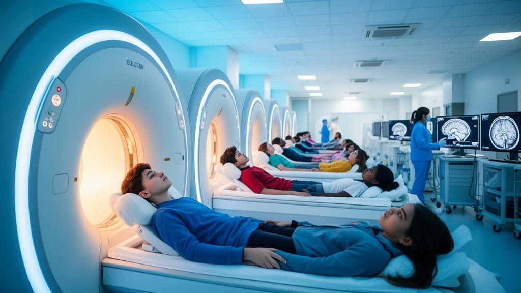
Researchers at the Perelman School of Medicine at the University of Pennsylvania have unveiled one of the most detailed pictures yet of how the adolescent brain matures, drawing on an extraordinary trove of magnetic resonance imaging data. The team processed more than 24,000 MRI scans from over 10,000 young people, producing a large, openly available neuroimaging resource that charts how the brain’s internal wiring changes through adolescence. Writing in the journal Neuron, the researchers describe remarkably consistent patterns of white matter development that emerged despite the scans having been collected at 21 different clinical sites using different scanner technologies and with varying image quality. The work was led by senior author Ted Satterthwaite, the McLure II Professor of Psychiatry and Behavioral Research and director of the Penn Lifespan Informatics and Neuroimaging Center, together with first author Steven Meisler, a postdoctoral fellow in the same laboratory.
The raw material for the project came from the Adolescent Brain Cognitive Development Study, or ABCD, the largest long-term study of brain development and child health in the United States, funded by the National Institutes of Health. Because ABCD follows thousands of children over multiple years, it collects repeated scans from the same participants at different points in time, which is essential for distinguishing true developmental change from individual differences between people. The Penn-led team took the more than 24,000 scans gathered from over 10,000 adolescents across those longitudinal visits and subjected them to a rigorous, standardized processing pipeline, transforming raw images into analyzable measures of brain structure. The result is a curated dataset known as the ABCD-BIDS Community Collection, which the researchers have released so that scientists anywhere in the world can study adolescent brain development without repeating the enormous technical effort of processing the images themselves.
At the heart of the study is a technique called diffusion MRI, which does not simply photograph the brain but probes its microstructure. By measuring how water molecules move through tissue, diffusion MRI reveals the organization of white matter, the bundles of nerve fibers that connect distant brain regions and allow them to communicate. Because water diffuses more easily along the length of an axon than across it, the direction and speed of water movement encode information about fiber density, coherence, and integrity. Adolescence is a critical period for this wiring: the long-range connections that support attention, learning, emotion regulation, and executive function continue to mature well into the teenage years, and disruptions in these trajectories have been linked to the emergence of psychiatric illness during this window of life.
Conventional analyses of diffusion MRI typically rely on relatively simple summary measures, such as fractional anisotropy, which captures the overall directionality of water diffusion in a region of white matter. These measures have been workhorses of developmental neuroscience for decades, but they are inherently limited, because they collapse complex microstructural information into a single number. The Penn team instead applied more advanced diffusion models that extract richer, multidimensional features of white matter organization. What they found was striking: these sophisticated measures uncovered developmental changes across adolescence that were far less apparent with traditional metrics. In other words, some aspects of teenage brain maturation have been hiding beneath the resolution of the field’s most commonly used tools.
The technical achievement underlying these findings should not be understated. When tens of thousands of scans are collected on different scanners, running different software versions, at dozens of sites spread across the country, the technical differences between images can dwarf the subtle biological changes scientists hope to measure. As Meisler explained, those site and scanner effects can be larger than the developmental signals themselves, threatening to swamp any genuine pattern. To overcome this, the researchers turned to advanced open-source image-processing and statistical methods developed at Penn specifically for large, multisite neuroimaging studies. These pipelines harmonize the data, statistically accounting for differences in acquisition and quality so that scans from different machines can be meaningfully compared.
The payoff from this harmonization was clear. After correcting for technical differences, the team examined how multiple features of white matter changed across adolescence and found widespread, highly consistent developmental patterns that held up across all 21 study locations. Satterthwaite emphasized that despite substantial variation in image quality across tens of thousands of scans acquired by different scanners, the patterns of white matter development were remarkably consistent. This consistency matters enormously for the credibility of large-scale developmental neuroscience, because inconsistent findings across sites have long been a weakness of multisite imaging studies. Demonstrating that a carefully harmonized pipeline can produce reproducible developmental trajectories at this scale offers reassurance that the biological signals are real and not artifacts of measurement.
Beyond the scientific findings themselves, a central ambition of the project was reproducibility. Large research consortia generate datasets of extraordinary scale, but the processing required to make those data scientifically useful is often difficult, sometimes impossible, for individual laboratories to reproduce on their own. Meisler noted that by releasing both the processed data and the open-source tools used to create them, the team hopes other researchers can build on this work rather than starting from scratch. This philosophy reflects a broader movement in neuroscience toward open science, in which shared data, shared code, and shared pipelines allow results to be verified, extended, and reanalyzed in ways that closed research simply cannot permit. The dataset is openly accessible, lowering the barrier for researchers worldwide who study adolescent development but lack the computing infrastructure or consortium membership to generate such resources independently.
The implications of the resource extend well beyond white matter biology. Researchers in the PennLINC laboratory are already using the collection to investigate how white matter development relates to environmental exposures and other aspects of adolescent health. Because the ABCD Study also tracks participants’ environments, experiences, and behaviors in fine detail, the processed imaging data can be linked to a rich web of developmental context, from socioeconomic factors to substance use to academic outcomes. This opens the door to asking precisely how the events of adolescence, both beneficial and harmful, shape the maturing brain’s connectivity during a period when it is especially plastic. The consistency of the developmental patterns uncovered here also provides a normative backdrop against which atypical development, and the early emergence of psychiatric conditions, can be measured.
For the field of developmental neuroscience, the study signals a methodological turning point. It shows that state-of-the-art diffusion MRI, combined with harmonization methods built for scale, can extract developmental signals from real-world, multisite data that conventional approaches may miss. It also demonstrates that the perceived trade-off between scale and quality, between the statistical power of thousands of participants and the precision of carefully controlled imaging, can be resolved with the right processing tools. As similar open collections are built for other populations and age ranges, researchers anticipate a future in which normative charts of brain development, analogous to the growth curves pediatricians use for height and weight, become a routine benchmark for identifying when a young person’s brain development deviates from the expected path.
The ABCD-BIDS Community Collection thus represents both a dataset and a statement about how twenty-first-century neuroscience should be done: collaboratively, transparently, and at scale. By pairing one of the largest longitudinal imaging collections ever assembled with openly documented, reproducible processing, the Penn-led team has converted tens of thousands of raw scans into a durable scientific infrastructure. The consistent white matter trajectories it reveals illuminate one of the most dynamic periods of human brain maturation, while the resource itself ensures that the next generation of discoveries about the adolescent brain will not have to be built from the ground up. The findings were published in Neuron, and the processed data are freely available to the research community.
Subject of Research: White matter development in the adolescent brain measured with diffusion MRI from a large multiscale longitudinal neuroimaging dataset
Article Title: Advanced MRI dataset maps adolescent brain development
Article References: Advanced MRI dataset maps adolescent brain development. (n.d.). Original publication
Image Credits: AI Generated
DOI: Not provided
Keywords: adolescent brain development, diffusion MRI, white matter, ABCD Study, neuroimaging, open science, brain connectivity, harmonization, Neuron, longitudinal study, University of Pennsylvania, data sharing
Cite Scienmag News
Cassandra Pierce. (October 18, 2026). Massive MRI collection reveals hidden patterns of adolescent brain wiring. Scienmag. https://scienmag.com/massive-mri-collection-reveals-hidden-patterns-of-adolescent-brain-wiring/
Cassandra Pierce. "Massive MRI collection reveals hidden patterns of adolescent brain wiring." Scienmag, 18 October 2026, https://scienmag.com/massive-mri-collection-reveals-hidden-patterns-of-adolescent-brain-wiring/. Accessed 18 October 2026.
Cassandra Pierce. "Massive MRI collection reveals hidden patterns of adolescent brain wiring." Scienmag. October 18, 2026. https://scienmag.com/massive-mri-collection-reveals-hidden-patterns-of-adolescent-brain-wiring/


