


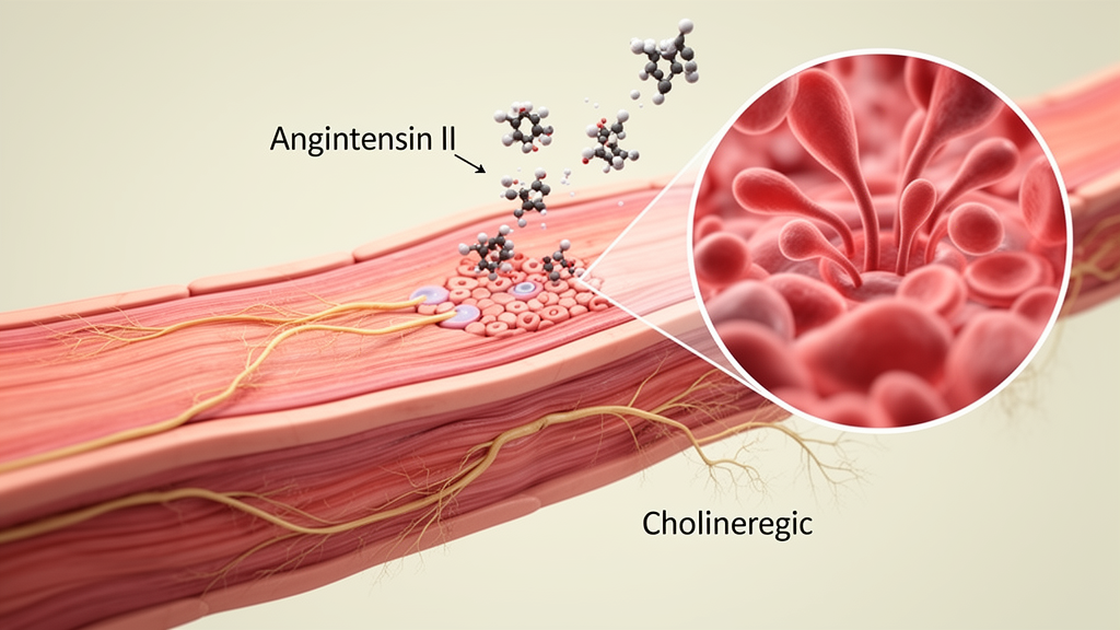
Diverticular disease affects millions of people worldwide, yet the cellular events that turn small pouches in the colonic wall into symptomatic, sometimes inflamed lesions remain stubbornly opaque. A research team from the Lithuanian University of Health Sciences in Kaunas, working with colleagues at the Sahlgrenska Academy in Gothenburg, has now uncovered a striking imbalance in the chemical control of colonic circular smooth muscle that may help explain how these pouches form in the first place. Their study, published in Frontiers in Physiology, shows that in patients with diverticular disease the circular muscle layer overreacts to angiotensin II, a hormone better known for constricting blood vessels, while simultaneously losing part of its responsiveness to acetylcholine, the chief excitatory neurotransmitter of the gut.
The circular muscle layer is the engine of colonic segmentation, the rhythmic pinching motion that divides the colon into compartments. Classic experiments dating back to the 1960s demonstrated that this segmentation generates localized pockets of high intraluminal pressure, and that the resulting pulsion force can push mucosa through weak points in the colonic wall, creating diverticula. Because functional abnormalities in diverticular disease concentrate in the circular layer while the longitudinal layer largely retains normal electrophysiological activity, understanding what drives circular muscle contraction is central to understanding the disease. Abnormal motility is thought to be an early pathogenic event that precedes the structural hallmarks, including thickening of the circular muscle and contracture of the taeniae coli.
To probe the chemical wiring of this layer, the investigators obtained full-thickness colonic specimens from 16 patients with diverticular disease and 16 controls undergoing colorectal surgery for cancer, all from macroscopically normal regions of the colon. Patients with irritable bowel syndrome, food intolerance, coeliac disease, Crohn’s disease, ulcerative colitis, arterial hypertension, or any medication known to affect gastrointestinal motility or the renin-angiotensin system were excluded, a design choice that isolates disease-related changes from drug-induced adaptations. The two groups were statistically comparable in age, sex, body mass index, and inflammatory markers, with the sigmoid colon the most common site of diverticula, consistent with decades of clinical observation.
The team first mapped where the two angiotensin II receptors reside. Immunofluorescence on 18-micrometer cryosections revealed a clean division of labor: the type 1 receptor, AT1R, was predominantly localized in colonic smooth muscle cells of both the circular and longitudinal layers, whereas the type 2 receptor, AT2R, was mainly detected in the myenteric plexus and in nerve fibers running between the muscle layers, with no detectable expression in smooth muscle cells. This split distribution matters because it suggests angiotensin II can act directly on the muscle machinery through AT1R while also engaging the enteric nervous system through AT2R, two routes with potentially different consequences for contractility.
Biochemical quantification then showed that the local renin-angiotensin system is genuinely activated in the diseased colon. Enzyme-linked immunosorbent assay measured angiotensin II concentrations in colonic muscular tissue of 529.7 picograms per milliliter in the diverticular disease group versus 321.2 picograms per milliliter in controls, a significant difference. Western blotting and quantitative real-time PCR added a receptor-level twist: AT2R protein expression was significantly higher in the diseased tissue, 4.58 versus 2.37 normalized units, and AT2R mRNA was also upregulated, while AT1R mRNA and protein expression did not differ significantly between groups. The enhanced AT2R signal likely reflects its neuronal pool, since the entire muscular layer, including the myenteric plexus, was analyzed together.
The functional experiments were performed with organ bath recordings, the gold standard for measuring isometric force in isolated muscle. Circular muscle strips roughly 2 by 10 millimeters were stretched to 1 gram, equilibrated for 45 minutes in warm, oxygenated Krebs solution, and then challenged with cumulative concentrations of angiotensin II ranging from 10 to the minus 10 to 10 to the minus 6 molar. The hormone produced concentration-dependent contractions with a half-maximal effective concentration of 3.25 times 10 to the minus 8 molar, and the maximal response was achieved at 10 to the minus 6 molar, the concentration used for all comparative experiments. Responses were expressed as a percentage of the contraction elicited by bethanechol, a stable muscarinic agonist applied at 10 to the minus 4 molar as an internal reference.
Here the results diverged sharply between the two chemical systems. Bethanechol-induced maximal contraction was significantly weaker in the diverticular disease group, with peak forces of 6.44 grams versus 8.14 grams in controls, and mean 5-minute forces of 4.02 versus 5.34 grams, indicating impaired cholinergic contractile function. Angiotensin II, by contrast, elicited significantly stronger contractions in the diseased tissue: the peak response reached 60.47 percent of the bethanechol reference in diverticular disease versus 40.91 percent in controls, and the mean 5-minute response was 51.51 versus 39.43 percent. The same hormone, in the same concentration, produced a fundamentally different reaction in muscle that had lost part of its acetylcholine responsiveness.
Pharmacological blockade clarified which receptor carries the signal. Losartan, a selective AT1R antagonist applied at 10 to the minus 7 molar, significantly reduced the mean 5-minute angiotensin II response in both groups, from 39.43 to 25.76 percent of the reference in controls and from 51.51 to 38.40 percent in diverticular disease. Yet even after AT1R blockade, both peak and mean responses remained significantly greater in the diseased tissue, 48.82 versus 29.98 percent at peak, suggesting reduced responsiveness to AT1R blockade in diverticular disease. Neither tetrodotoxin, which silences neural action potentials, nor PD123319, the AT2R antagonist, significantly affected the contractions, indicating that angiotensin II acts primarily through direct activation of smooth muscle rather than through enteric neural pathways, and that AT2R plays no detectable role in the contractile response under these conditions.
The authors acknowledge limitations, including that losartan-treated and untreated responses came from independent tissue samples rather than paired preparations, and that their full-thickness analyses could not distinguish membrane-associated from intracellular receptor pools, leaving open whether AT1R trafficking is altered. Previous work in rat smooth muscle cells has shown that high angiotensin II concentrations induce AT1R internalization, a physiological trafficking mechanism that may limit excessive receptor activation. Because the patients had not received angiotensin receptor blockers or ACE inhibitors, the enhanced contractile responses most likely reflect disease-related activation of the local renin-angiotensin system rather than adaptations to chronic pharmacological blockade.
Taken together, the findings sketch a regulatory shift in the diseased colon: as cholinergic control of the circular muscle weakens, angiotensin II becomes a more prominent contractile regulator, potentially promoting the abnormal circular muscle activity and elevated intraluminal pressure that drive diverticulum formation. The distinct muscular and neural localization of AT1R and AT2R implies differential roles for angiotensin signaling at these two levels, and the upregulated AT2R in enteric neurons hints at a neural dimension that the current contractile assays could not capture. Whether drugs that temper local angiotensin signaling could one day ease the motility disturbances of diverticular disease remains speculative, but this study provides the first direct evidence in human tissue that the hormone’s contractile influence is amplified precisely where the disease does its damage.
Subject of Research: Physiology
Article Title: Altered contractile responses to angiotensin II and cholinergic stimulation in colonic circular smooth muscle from patients with diverticular disease
Article References: Malinauskas, M., Žilinskas, J., Stukas, D., Rysevaite-Kyguoliene, K., Nekrosius, D., Paužas, H., Macas, A., & Casselbrant, A. (2026). Altered contractile responses to angiotensin II and cholinergic stimulation in colonic circular smooth muscle from patients with diverticular disease. Frontiers in Physiology, 17, Article 1991333. https://doi.org/10.3389/fphys.2026.1991333
Image Credits: AI Generated
DOI: 10.3389/fphys.2026.1991333
Keywords: diverticular disease, angiotensin II, colon, smooth muscle, renin-angiotensin system, AT1 receptor, AT2 receptor, cholinergic, gastrointestinal physiology, organ bath, Altered, contractile
Cite Scienmag News
Drew Townsend. (October 17, 2026). Colonic Muscle in Diverticular Disease Overreacts to Angiotensin II While Losing Cholinergic Drive. Scienmag. https://scienmag.com/colonic-muscle-in-diverticular-disease-overreacts-to-angiotensin-ii-while-losing-cholinergic-drive/
Drew Townsend. "Colonic Muscle in Diverticular Disease Overreacts to Angiotensin II While Losing Cholinergic Drive." Scienmag, 17 October 2026, https://scienmag.com/colonic-muscle-in-diverticular-disease-overreacts-to-angiotensin-ii-while-losing-cholinergic-drive/. Accessed 17 October 2026.
Drew Townsend. "Colonic Muscle in Diverticular Disease Overreacts to Angiotensin II While Losing Cholinergic Drive." Scienmag. October 17, 2026. https://scienmag.com/colonic-muscle-in-diverticular-disease-overreacts-to-angiotensin-ii-while-losing-cholinergic-drive/


