


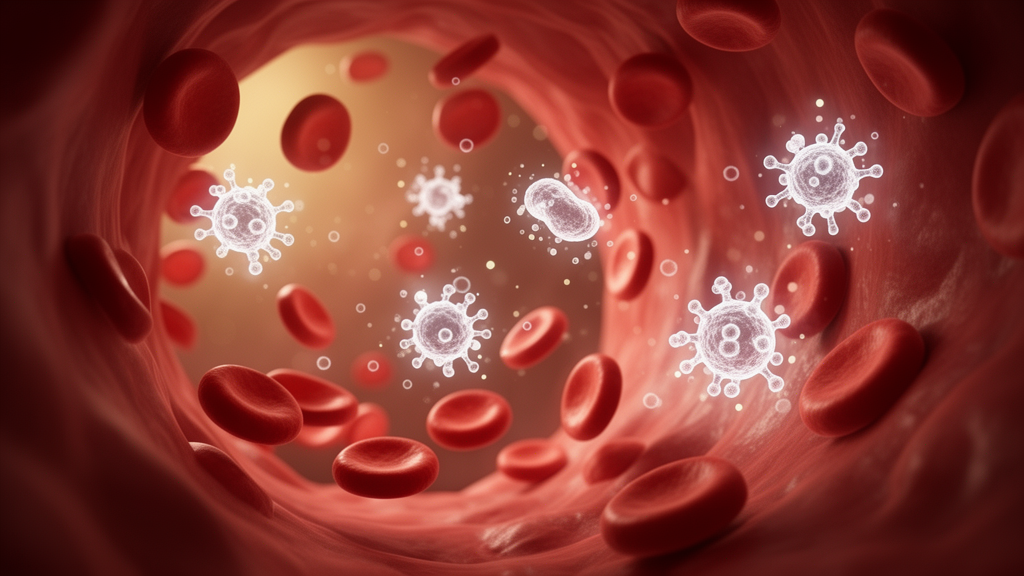
Chronic exposure to high levels of cortisol, the body’s principal stress hormone, leaves a measurable fingerprint on the immune system. A new retrospective cross-sectional study from researchers in Istanbul, Türkiye, now maps that fingerprint in detail, comparing the white blood cell profiles of patients with overt Cushing syndrome, patients with a milder form of hormone excess known as mild autonomous cortisol secretion, and patients carrying nonfunctioning adrenal tumors that produce no excess hormone at all. The findings, published in BMC Endocrine Disorders, show that only full-blown cortisol excess is accompanied by a distinct expansion of the myeloid arm of the immune system, while the milder condition looks essentially indistinguishable from hormone-silent tumors on routine blood counts.
The research team, led by endocrinologists at Basaksehir Cam and Sakura City Hospital affiliated with the University of Health Sciences, analyzed records from 252 patients. The cohort comprised 132 patients with overt Cushing syndrome, subdivided into 75 with Cushing disease, where a pituitary tumor drives excess adrenocorticotropic hormone, and 57 with adrenal Cushing syndrome, where the adrenal gland itself overproduces cortisol. A further 68 patients had mild autonomous cortisol secretion, often abbreviated MACS, a subtle biochemical abnormality frequently detected in adrenal incidentalomas, and 52 patients had nonfunctioning adrenal tumors, or NFAT, which secrete no excess hormone. The investigators compared standard complete blood count parameters across these groups, including total white blood cell count and the individual counts of neutrophils, monocytes, lymphocytes, eosinophils, and basophils, together with two widely used derived indices, the neutrophil-to-lymphocyte ratio and the monocyte-to-lymphocyte ratio.
Cortisol is a potent immunomodulatory hormone, and its chronic excess has long been known to reshape circulating leukocyte populations. Glucocorticoids promote the release of mature neutrophils from the bone marrow, delay their apoptosis, and simultaneously drive the redistribution and death of lymphocytes, producing the classic picture of neutrophilia with relative lymphopenia seen in states of glucocorticoid excess. What has remained uncertain is whether the low-grade, often subclinical cortisol elevation of MACS produces hematological changes that are detectable and comparable to those of overt disease. Resolving this question matters clinically, because MACS is increasingly diagnosed as adrenal imaging becomes more common, yet its systemic consequences remain debated.
The results were clear-cut at the extremes. Total white blood cell count, neutrophil count, monocyte count, and the monocyte-to-lymphocyte ratio were all significantly higher in patients with overt Cushing syndrome than in either the MACS group or the NFAT group, with p values at or below 0.002. The neutrophil-to-lymphocyte ratio, a marker frequently studied in inflammatory and endocrine disease, was significantly elevated in overt Cushing syndrome only in comparison with MACS once the analysis was corrected for multiple comparisons using the Bonferroni method. This pattern indicates that the myeloid expansion characteristic of glucocorticoid excess, with more neutrophils and monocytes relative to lymphocytes, is a feature of overt disease rather than of the milder biochemical state.
In striking contrast, no significant differences emerged between the MACS and NFAT groups on any individual parameter, with all p values exceeding 0.05. The effect sizes separating these two groups were uniformly small, with Cohen’s d values ranging from 0.02 to 0.17, a range conventionally interpreted as negligible. The authors note that any true difference between mild autonomous cortisol secretion and nonfunctioning tumors is likely to be small, although they caution that subtle effects cannot be entirely excluded by a study of this size. In practical terms, a routine blood count cannot currently distinguish a patient with mild cortisol autonomy from one with a hormone-silent adrenal mass.
Beyond group comparisons, the study examined how leukocyte parameters track with biochemical measures of cortisol burden. Neutrophil count, monocyte count, the neutrophil-to-lymphocyte ratio, and the monocyte-to-lymphocyte ratio each correlated positively with multiple cortisol markers, which in this clinical context typically include urinary free cortisol, late-night salivary cortisol, and the dexamethasone suppression test. This dose-response relationship strengthens the biological plausibility of the findings: the more cortisol circulating, the more pronounced the shift toward myeloid cells in the peripheral blood. It also suggests that these inexpensive, routinely measured indices could serve as accessible proxies for the immunological impact of cortisol excess.
To move beyond univariate comparisons, the researchers performed an exploratory linear discriminant analysis, a multivariate statistical technique that seeks linear combinations of variables best separating predefined groups. The analysis aimed to characterize the overall leukocyte profiles of Cushing disease, adrenal Cushing syndrome, MACS, and NFAT. On the primary discriminant axis, basophil count emerged as the parameter with the highest normalized loading. However, the authors themselves flag an important statistical caveat: because basophil counts are small in absolute magnitude, their loadings can be inflated by scale-related artifacts rather than reflecting a genuine biological signal. This transparency is a notable feature of the paper, which consistently frames its multivariate results as exploratory.
Indeed, the authors are explicit that the classification performance of the discriminant model was modest and not validated in an independent cohort. They emphasize that these findings are hypothesis-generating and should not be interpreted as evidence of a validated diagnostic tool or of a pathophysiological continuum linking mild and overt cortisol excess. This restraint matters in a field where machine-learning-style analyses of routine laboratory data are increasingly promoted as diagnostic shortcuts. The study instead delivers a more measured message: overt hypercortisolism leaves a clear myeloid signature in the blood, but mild autonomous cortisol secretion, at least as captured by standard hematological parameters, does not.
The clinical implications cut in two directions. For patients with overt Cushing syndrome, the correlation between myeloid indices and cortisol burden adds to the evidence that chronic glucocorticoid excess produces measurable systemic inflammation-like changes, which may be relevant to the elevated cardiovascular and infectious risk seen in these patients. For the growing population of patients found to have adrenal incidentalomas, the absence of hematological differences between MACS and NFAT suggests that routine blood counts offer no added diagnostic value in characterizing the functional status of such tumors, and that established endocrine testing remains essential.
As a single-center retrospective cross-sectional study, the work has inherent limitations, including the possibility of selection bias and the inability to establish causality or track changes over time within individual patients. Nevertheless, by systematically comparing four clinically distinct adrenal and pituitary states within a single cohort, the study provides one of the clearest pictures to date of where, on the spectrum of cortisol excess, the immune system begins to visibly shift. The answer, apparently, is not at the mild end. Future prospective studies with larger MACS cohorts and longitudinal sampling will be needed to determine whether subtle immune changes emerge over time, and whether any leukocyte signature could eventually complement, rather than replace, the biochemical tests that currently define this enigmatic condition.
Subject of Research: Peripheral leukocyte profiles across clinical forms of hypercortisolism and nonfunctioning adrenal tumors
Article Title: Peripheral leukocyte profiles in different clinical forms of hypercortisolism and nonfunctioning adrenal tumors: a retrospective cross-sectional study
Article References: Burhan, S., Ozkahraman, A. U., Korkmaz Yilmaz, M., Akbulut, S., Cicek, M. Z., Niyazoglu, M., & Hatipoglu, E. (2026). Peripheral leukocyte profiles in different clinical forms of hypercortisolism and nonfunctioning adrenal tumors: a retrospective cross-sectional study. BMC Endocrine Disorders. https://doi.org/10.1186/s12902-026-02668-2
Image Credits: AI Generated
DOI: 10.1186/s12902-026-02668-2
Keywords: Cushing syndrome, mild autonomous cortisol secretion, adrenal incidentaloma, leukocytes, neutrophil-to-lymphocyte ratio, monocyte-to-lymphocyte ratio, cortisol, endocrinology, hematology, linear discriminant analysis, biomarkers, nonfunctioning adrenal tumor
Cite Scienmag News
Kristina Jarvis. (October 16, 2026). Blood Cell Patterns Reveal How Excess Cortisol Reshapes the Immune System. Scienmag. https://scienmag.com/blood-cell-patterns-reveal-how-excess-cortisol-reshapes-the-immune-system/
Kristina Jarvis. "Blood Cell Patterns Reveal How Excess Cortisol Reshapes the Immune System." Scienmag, 16 October 2026, https://scienmag.com/blood-cell-patterns-reveal-how-excess-cortisol-reshapes-the-immune-system/. Accessed 16 October 2026.
Kristina Jarvis. "Blood Cell Patterns Reveal How Excess Cortisol Reshapes the Immune System." Scienmag. October 16, 2026. https://scienmag.com/blood-cell-patterns-reveal-how-excess-cortisol-reshapes-the-immune-system/


