


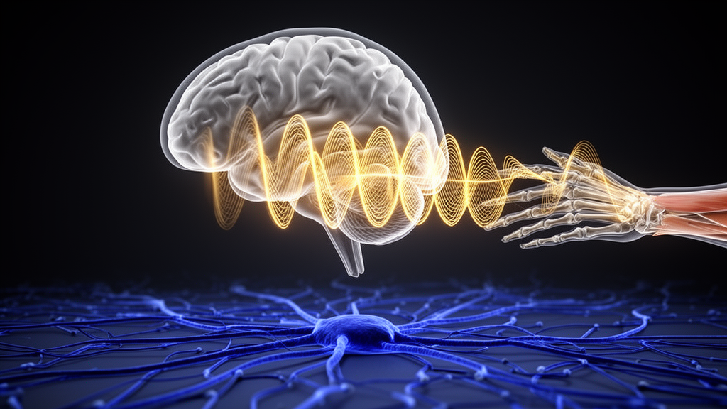
Deep inside the brain, a metronome is not always needed for rhythm. In a rare study of the human subcortex, researchers have recorded electrical activity directly from the brain’s deep motor circuits while a teenager with dystonia tapped out a beat, and the recordings show that networks long associated with movement disorders are also exquisitely tuned to the passage of time. The findings, published in the journal Dystonia, offer some of the most direct human evidence yet that both the basal ganglia and the thalamic relay stations for cerebellar signals participate in coordinating rhythmic movement, and they may reshape how neurosurgeons think about targeting deep brain stimulation.
The study took advantage of an unusual clinical circumstance. A 14-year-old boy with generalized secondary dystonia was undergoing staged implantation of deep brain stimulation (DBS) leads as part of his treatment at Children’s Hospital of Orange County. Before the therapeutic electrodes were finalized, temporary stereoelectroencephalography (sEEG) leads were placed in candidate DBS targets, giving the research team a window into brain regions that almost never lend themselves to direct recording in humans. With informed consent from his guardians and approval from the institutional review boards, the team turned a surgical procedure into a scientific experiment on motor timing.
Ten depth leads were implanted bilaterally, reaching the globus pallidus internus (GPi) and subthalamic nucleus (STN) within the basal ganglia, the ventral oralis (Vo) and centromedian (CM) nuclei of the thalamus, and two thalamic nuclei that receive direct projections from the cerebellum: the ventral intermediate nucleus (VIM) and the centrolateral nucleus (CL). Each lead carried low-impedance macro-contacts and high-impedance microwire electrodes, with micro-contact signals sampled at roughly 24 kilohertz and preamplified at the connector to suppress noise and motion artifacts. From these channels, the researchers extracted local field potentials (LFPs), the summed electrical chatter of local neural populations, filtered between 0.05 and 30 hertz.
While the electrodes were in place, the participant performed a classic two-phase timing task drawn from decades of cognitive neuroscience research. In each 18-second trial, an auditory metronome delivered tones every 600 milliseconds for the first 6 seconds, and the boy moved a joystick in sync with the beeps. Then the metronome fell silent. For the remaining 12 seconds, he had to keep the rhythm going from memory alone. This split between synchronization and continuation is the standard way researchers separate externally cued timing, which depends on sensory prediction, from internally generated timing, which relies on the brain’s own clock.
The behavioral data already told an interesting story. During the first three intervals after the metronome started, his movements lagged behind the tones, but he quickly flipped into anticipation, with responses beginning to lead the auditory cues by a stable margin. More striking, he did not simply obey the metronome. His average interval was 557 milliseconds in the cued phase and sped up to 536 milliseconds when the tones stopped, both faster than the prescribed 600 milliseconds. The fact that he carried his own tempo from the cued phase into the continuation phase suggests he was relying on an endogenous timing signal rather than passively entraining to the external beat. His timing variability was actually lower without the metronome, and his Weber fraction, a normalized measure of temporal precision, was 9.45 percent in the continuation phase, somewhat above the roughly 5 percent typical of young adults, a difference the authors attribute to his age, his neurological condition, and the response device.
The neural recordings were the heart of the study. When the researchers averaged LFPs time-locked to joystick movement onset across trials, they found strong, rhythmic modulations in every recorded region, including the cerebellar-recipient VIM and CL. These were not diffuse fluctuations; the signals were phase-locked to the movements, indicating functional coupling between neural activity and motor output at the level of individual actions. Crucially, the amplitude of these time-locked responses was significantly stronger during the synchronization phase than during the continuation phase in all regions, after statistical correction for multiple comparisons. The researchers suggest several interpretations: the larger cued-phase responses may reflect neural reactions to the tones, heightened responsiveness when movement is paired with an auditory stimulus, or activity that monitors the phase relationship between what is heard and what is done.
Time-frequency analysis sharpened the picture further. Scalograms of the joystick signal showed sustained power at approximately 1.8 hertz, matching the participant’s own movement rate, throughout both task phases. The LFPs from the VIM and CL displayed oscillatory power at the same frequency, present while the metronome sounded and remaining stable after it stopped. Across all recorded regions, the power spectra during the task showed a prominent peak near the metronome frequency of 1.667 hertz, standing out clearly against resting baselines recorded before the task began. In other words, the deep motor circuits were oscillating at precisely the frequency of the behavior, in both the externally cued and the internally driven halves of the task. This frequency-specific entrainment across basal ganglia and cerebellothalamic circuits supports the idea that rhythmic timing emerges from coordinated activity distributed across these loops rather than from a single timing center.
The authors are careful about what the oscillations do and do not prove. They note that the rhythmicity of the neural responses likely reflects the iterative operation of an internal interval-timing mechanism rather than an autonomous oscillator ticking away on its own. They also acknowledge a classic confound: in this task, internal timing, motor execution, and sensory feedback are temporally aligned, so the recorded modulations could serve any or all of those functions. Interestingly, the Vo and VIM nuclei showed movement-related oscillations at slightly lower frequencies than other regions, hinting that their distinct inputs from cerebellum and basal ganglia may support an integration process for predictive control over extended intervals. Disentangling these roles, the researchers write, will require task designs that separate the components and, ideally, direct recordings from the cerebellum itself to trace where timing signals originate and how they propagate.
The clinical stakes are considerable. Dystonia is clinically defined by abnormal co-contraction and twisting postures rather than by rhythmic movement, but the study’s authors argue that impaired temporal coordination within subcortical circuits may contribute to that disorganization. If cerebellothalamic and basal ganglia pathways each carry precisely timed, behaviorally locked signals, then the choice of DBS target becomes more than a matter of anatomy; it becomes a question of which timing circuit to modulate. The VIM, which receives excitatory input from the cerebellar dentate nucleus and has been implicated in event timing, and the CL, another cerebellar recipient, emerge as candidate relay sites for timing-related signals within a distributed motor network. Understanding these dynamics could help clinicians tune stimulation parameters to restore temporal stability, not just suppress symptoms.
The researchers also lay out the limits of the work with unusual candor. The dataset comes from a single participant, which constrains generalizability, although several features of the design mitigate concerns that dystonia itself drove the results. The task required only small, controlled joystick movements that were largely unaffected by dystonic posturing, and the analyses focused on low-frequency dynamics around 1 to 2 hertz, bands unlikely to be dominated by dystonia-related activity. The team proposes extending the approach to multi-subject datasets, testing trial-by-trial correlations between LFP power and behavioral precision, and applying cross-regional coherence analysis and computational modeling to separate the contributions of sensory-guided and internally generated timing. For now, the study stands as a vivid demonstration that when the brain keeps a beat, the echo of that beat can be read directly from the circuits that deep brain stimulation is built to reach, and that time itself may be a measurable currency flowing between the basal ganglia and the cerebellar thalamus.
Subject of Research: Neural mechanisms of motor timing in basal ganglia and cerebellothalamic circuits in dystonia
Article Title: Behaviorally coupled oscillations reveal distinct timing roles of basal ganglia and cerebellothalamic circuits in dystonia
Article References: Behaviorally coupled oscillations reveal distinct timing roles of basal ganglia and cerebellothalamic circuits in dystonia. (n.d.). https://doi.org/10.3389/dyst.2025.15390
Image Credits: AI Generated
Keywords: dystonia, deep brain stimulation, motor timing, basal ganglia, cerebellum, thalamus, local field potentials, stereoelectroencephalography, neural oscillations, rhythmic movement, ventral intermediate nucleus, pediatric neurosurgery
Cite Scienmag News
Cassandra Pierce. (October 16, 2026). Brain Rhythms Locked to Movement Reveal How Deep Circuits Keep Time in Dystonia. Scienmag. https://scienmag.com/brain-rhythms-locked-to-movement-reveal-how-deep-circuits-keep-time-in-dystonia/
Cassandra Pierce. "Brain Rhythms Locked to Movement Reveal How Deep Circuits Keep Time in Dystonia." Scienmag, 16 October 2026, https://scienmag.com/brain-rhythms-locked-to-movement-reveal-how-deep-circuits-keep-time-in-dystonia/. Accessed 16 October 2026.
Cassandra Pierce. "Brain Rhythms Locked to Movement Reveal How Deep Circuits Keep Time in Dystonia." Scienmag. October 16, 2026. https://scienmag.com/brain-rhythms-locked-to-movement-reveal-how-deep-circuits-keep-time-in-dystonia/


