


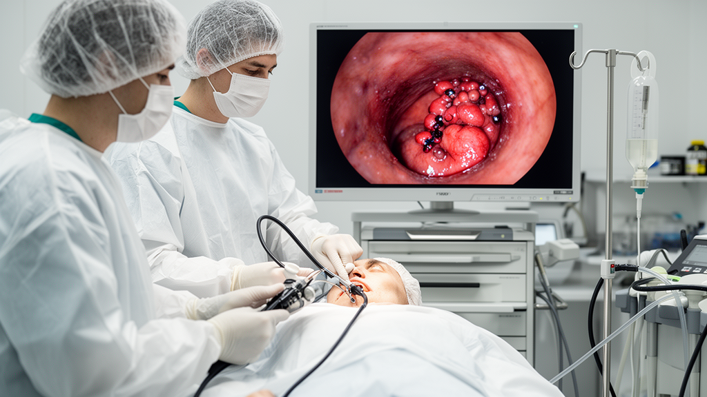
A 59-year-old man in Beijing who had bled from his bowels since birth — at one point dropping to a hemoglobin level of just 40–50 g/L and requiring repeated transfusions — has become the first reported patient with Klippel–Trénaunay syndrome and extensive anorectal venous malformations to be successfully treated with a combination of endoscopic lauromacrogol sclerotherapy and band ligation. The case, published in Frontiers in Medicine by physicians at Peking Union Medical College Hospital, offers a rare glimpse into how one of medicine’s most obscure vascular disorders can be managed without resorting to drastic surgery.
Klippel–Trénaunay syndrome (KTS) affects roughly one in 100,000 people worldwide. It is a congenital mesodermal disorder defined by a classic triad: varicose veins, capillary malformations (often visible as port-wine stains), and bone or soft-tissue overgrowth of an affected limb. Crucially, KTS is distinguished from other overgrowth syndromes by the presence of slow-flow vascular malformations and the complete absence of arteriovenous malformations — a distinction that carries enormous clinical weight when deciding how to intervene.
At the molecular level, KTS belongs to what researchers now call the PIK3CA-related overgrowth spectrum, or PROS. Somatic mosaic mutations in the PIK3CA gene — which encodes the p110α catalytic subunit of the PI3K lipid kinase — cause structural alterations that prevent p110α from binding to its p85α regulatory subunit, the very protein that normally keeps its activity in check. The result is hyperactivation of the PI3K-AKT-mTOR pathway, driving dysregulated cell proliferation and the segmental overgrowth of blood vessels, soft tissue, and bone that defines the condition. Despite PIK3CA’s frequent role in cancer biology, these variants do not appear to increase malignancy risk in PROS patients.
For the Beijing patient, the disease had quietly escalated over decades. Physical examination revealed hypertrophy of the right buttock and lower limb, multiple subcutaneous varicose veins studded with calcified nodules, penile varicosities, and splenomegaly. MRI demonstrated venous malformation-like changes in the rectal and sigmoid colonic walls with secondary luminal stenosis. Contrast-enhanced CT showed rectal and sigmoid wall thickening studded with multiple calcifications — phleboliths, the telltale calcified thrombi that form inside slow-flowing venous blood — along with calcifications in the bilateral adrenal glands, pelvis, and scrotum, and splenic hemangiomas measuring up to 4.5 by 5.0 centimeters.
Colonoscopy proved decisive. It revealed a circumferential mucosal elevation with bluish discoloration extending from 15 centimeters above the anal verge, with active bleeding at 12 centimeters and clusters of smooth, grape-like nodular elevations at 5 centimeters. Endorectal ultrasound showed a thickened, hypoechoic submucosal layer with fine honeycomb-like changes and Doppler blood-flow signals — imaging hallmarks of venous malformations. Because biopsy of such lesions can trigger life-threatening hemorrhage, endoscopy serves a dual role: it identifies and maps the lesions while simultaneously warning clinicians against the one maneuver most likely to cause catastrophe.
After multidisciplinary team discussion, and with surgical resection ruled out because it would have precluded sphincter preservation, the team opted for local endoscopic therapy. Using a 23-gauge injection needle, they delivered 1–1.5 milliliters of 1% lauromacrogol per site — a total of 30 milliliters — at multiple points around the bleeding site and at the base of the venous malformations. Lauromacrogol, a polidocanol-like sclerosant, works by inducing endothelial injury, thrombosis, and eventual luminal occlusion through chemical stimulation. Immediately afterward, six bands were deployed in a spiral configuration around the bleeding site, targeting markedly elevated and erythematous areas at high risk of rebleeding, mechanically interrupting lesional blood flow.
Immediate hemostasis was achieved, with no procedure-related complications. At the 3-month follow-up, the patient’s hemoglobin had climbed to 105 g/L. By 6 months, he reported no obvious melena or hematochezia, and intermittent monitoring showed hemoglobin levels fluctuating steadily between 100 and 110 g/L. The team declined systemic medical therapy — citing cost, drug accessibility, and potential adverse effects — and the patient himself declined splenectomy, underscoring how patient preference and quality of life shape treatment decisions in rare, incurable conditions.
The authors emphasize that KTS remains incurable and that treatment is predominantly conservative and symptomatic, aimed at enhancing quality of life rather than curing the underlying disorder. Most previously reported cases of KTS-related gastrointestinal bleeding have been managed with partial bowel resection or conservative measures. Endoscopic approaches — sclerotherapy and argon laser coagulation — have been documented only in isolated case reports, though prior patients treated with polidocanol, ethanol, or argon laser coagulation have shown favorable bleeding control without reported complications. What makes this case notable is the specific combination of lauromacrogol injection and band ligation, an approach that has rarely, if ever, been reported for this indication.
One caution looms. The venous drainage of the upper rectum and sigmoid colon flows into the inferior mesenteric vein, which belongs to the portal venous system. The authors raise the concern that sclerosants and the thrombi they induce could migrate into the portal circulation, potentially triggering or worsening portal vein thrombosis and portal hypertension — which could, in turn, exacerbate the very malformations being treated. While this hypothesis requires further clinical confirmation, it implies that prolonged close monitoring is necessary after sclerotherapy, especially in patients with existing portal hypertension.
Meanwhile, targeted therapies are emerging from the molecular understanding of the disease. In 2022, alpelisib — a p110α inhibitor — became the first medical therapeutic drug approved by the US Food and Drug Administration for PROS, with efficacy demonstrated in the retrospective EPIK-P1 study, though lifelong therapy may be required. Low-dose sirolimus, an allosteric mTOR inhibitor, has shown moderate reductions in overgrowth but carries a relatively pronounced adverse-effect profile that demands individualized risk–benefit assessment. The ongoing VASE multicenter phase 3 trial has reported that individualized, intermittent sirolimus regimens can effectively control symptoms in PIK3CA-related venous malformations while significantly reducing adverse events. For now, however, the Beijing case suggests that for patients whose bleeding is driven by low rectal lesions unsuitable for sphincter-preserving surgery, the endoscope itself may be the most powerful tool available — provided clinicians recognize the syndrome early enough to use it safely.
Subject of Research: Endoscopic sclerotherapy and band ligation for gastrointestinal bleeding in Klippel–Trénaunay syndrome
Article Title: Endoscopic therapy for gastrointestinal bleeding in Klippel–Trénaunay syndrome: a case report and literature review
Article References: Zhang, R., Shi, W., Wu, X., Yang, A., & Chen, Y. (2026). Endoscopic therapy for gastrointestinal bleeding in Klippel–Trénaunay syndrome: a case report and literature review. Frontiers in Medicine, 13, Article 1960274. https://doi.org/10.3389/fmed.2026.1960274
Image Credits: AI Generated
DOI: 10.3389/fmed.2026.1960274
Keywords: Klippel–Trénaunay syndrome, vascular malformations, endoscopy, sclerotherapy, PIK3CA, rare disease, gastrointestinal bleeding, Endoscopic, therapy, gastrointestinal, bleeding, Klippel
Cite Scienmag News
Juliet Wilcox. (October 16, 2026). Doctors Stop Rare Genetic Bleeding Disorder With Endoscopic Sclerotherapy and Band Ligation. Scienmag. https://scienmag.com/doctors-stop-rare-genetic-bleeding-disorder-with-endoscopic-sclerotherapy-and-band-ligation/
Juliet Wilcox. "Doctors Stop Rare Genetic Bleeding Disorder With Endoscopic Sclerotherapy and Band Ligation." Scienmag, 16 October 2026, https://scienmag.com/doctors-stop-rare-genetic-bleeding-disorder-with-endoscopic-sclerotherapy-and-band-ligation/. Accessed 16 October 2026.
Juliet Wilcox. "Doctors Stop Rare Genetic Bleeding Disorder With Endoscopic Sclerotherapy and Band Ligation." Scienmag. October 16, 2026. https://scienmag.com/doctors-stop-rare-genetic-bleeding-disorder-with-endoscopic-sclerotherapy-and-band-ligation/


