


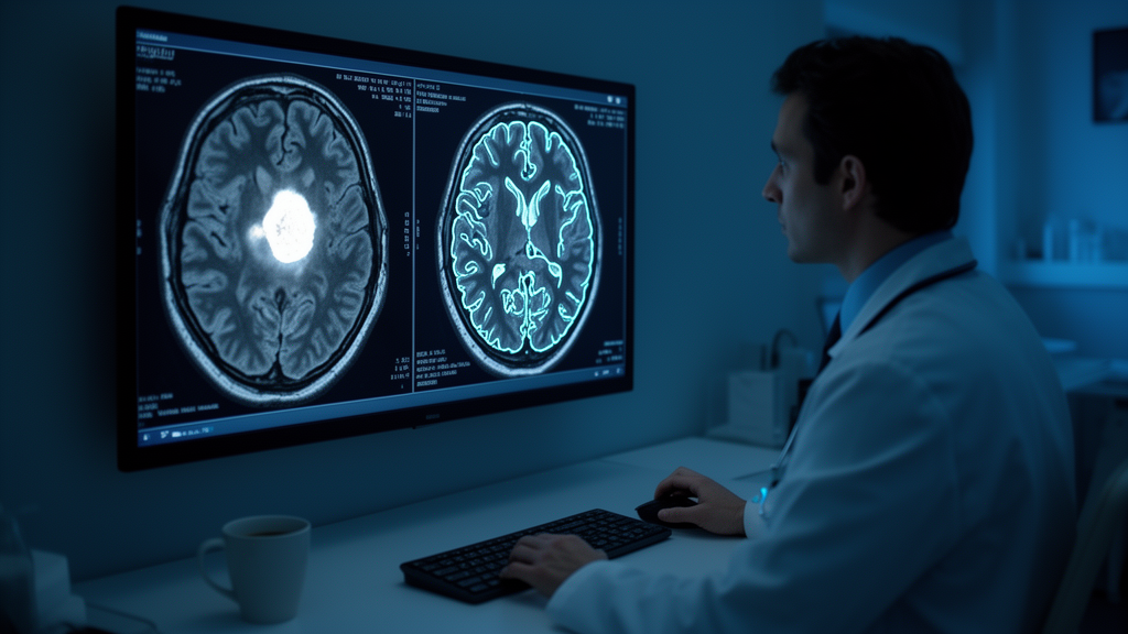
A team of researchers has unveiled a deep learning system that can look at a brain MRI scan and, in a fraction of a second, do two jobs at once: decide whether the scan shows any abnormality at all, and if it does, name the type of tumor present. The framework, described in the journal Discover Artificial Intelligence, is built on a modified version of the DenseNet121 convolutional neural network and pairs its predictions with heatmaps that show clinicians exactly where the model was looking. In an era when artificial intelligence in medicine is often criticized as an inscrutable black box, the study makes a deliberate push toward transparency, packaging accuracy and explanation inside a working desktop application.
The clinical stakes are considerable. Brain tumors produce symptoms that vary wildly depending on their type, grade, and location, ranging from headaches and seizures to motor impairment, sensory loss, and personality changes. Despite decades of progress in treatment, the five-year survival rate for malignant central nervous system tumors in the United States remains just 35.7 percent, a figure that underscores how much depends on early and accurate characterization. Radiologists reading MRI scans must first determine whether anything is wrong and then distinguish between tumor types that can look deceptively similar on T1-weighted images, particularly gliomas and meningiomas, which share overlapping morphological features that challenge even experienced eyes.
The researchers, led by Yasir Hussein Shakir with colleagues across institutions in Malaysia, Iraq, and Oman, identified four recurring weaknesses in the existing literature. Most published models handle either binary screening or multiclass classification, but not both, forcing hospitals to maintain and deploy two separate pipelines. Few models offer any visual explanation for their predictions, a roadblock to clinical trust. Accurate models rarely ship with usable software that a radiologist could actually integrate into a workflow. And nearly all prior studies validate on a single dataset, leaving generalizability an open question. The new system, called Modified DenseNet121, was designed to address all four gaps simultaneously.
Architecturally, the model starts from DenseNet121, a network whose defining feature is dense connectivity: every layer concatenates its feature maps with those of all preceding layers, enabling extensive feature reuse. The team chose this backbone because it carries fewer parameters than comparable ResNet designs and is less prone to vanishing or exploding gradients when fine-tuned on relatively small medical image collections. They stripped the ImageNet-pretrained network’s top classification layers, applied global average pooling to obtain a 1024-dimensional feature vector, and then attached a custom regularization stack: a dropout layer with a rate of 0.50, a 128-unit dense layer with ReLU activation for feature compression, and batch normalization to stabilize activations across training batches.
From that shared representation, two output branches diverge. The first is a single-unit sigmoid layer that predicts the probability that a scan is abnormal. The second is a three-unit softmax layer that assigns probabilities to glioma, meningioma, and pituitary tumors. A clever labeling strategy keeps the two tasks from interfering: every image contributes to the binary loss, but only abnormal images contribute to the multiclass loss, enforced through a binary mask that zeroes out the multiclass loss for healthy scans. This masking prevents the model from being misled by meaningless tumor labels on healthy subjects while allowing the shared backbone to learn robust representations for both tasks. The combined loss was optimized with the Adamax optimizer at a learning rate of 0.0001, a batch size of 32, and early stopping after ten epochs without validation improvement.
The benchmarking was unusually thorough. The team trained and evaluated the model against six competing architectures, including the unmodified DenseNet121 baseline, ConvNeXtSmall, ConvNeXtTiny, EfficientNetB0, NASNetMobile, and Xception, all pretrained on ImageNet and fine-tuned under identical preprocessing and evaluation conditions. Two large public MRI datasets served as the testbeds: a Kaggle collection of 7,023 T1-weighted contrast-enhanced scans and a Mendeley collection of 12,064 scans, both balanced across glioma, meningioma, pituitary, and normal classes. On the first dataset, Modified DenseNet121 achieved 99.85 percent binary accuracy and 99.00 percent multiclass accuracy, the best multiclass result overall, edging out the baseline by 0.28 percent. On the second, it reached 99.62 percent binary and 99.00 percent multiclass accuracy, statistically comparable to the baseline while retaining the advantage of a single dual-output framework.
Perhaps the most striking result came from a zero-shot evaluation on a third, entirely unseen dataset of 3,264 images. The model, trained on the merged 19,087 images from the first two datasets, achieved 99.75 percent binary accuracy and 99.20 percent multiclass accuracy without any additional fine-tuning, with bootstrap resampling over 1,000 iterations yielding narrow 95 percent confidence intervals. The authors are careful, however, to interpret this conservatively: because no dedicated image-level overlap analysis was performed between the public datasets, the result demonstrates external transfer performance rather than definitive proof of complete dataset independence. An ablation study further showed that while individual regularization components did not uniformly help, the full configuration delivered the best overall trade-off, with dropout removal causing the largest multiclass accuracy drop.
Interpretability comes through Gradient-weighted Class Activation Mapping, or Grad-CAM, a technique that computes the gradients of the predicted class score with respect to the feature maps of the network’s final convolutional block. Those gradients are globally average-pooled to produce importance weights for each feature map, which are then combined into a coarse localization map, resized to the original image resolution, and overlaid on the MRI with a jet colormap at 40 percent transparency. When a scan is classified as normal, no heatmap is generated; when abnormal, the heatmap highlights the tumor-relevant regions that drove the decision. In the published examples, the activations land where radiologists would expect: on an extra-axial hemispheric mass for a meningioma case, on a large intra-axial frontal-parietal lesion for a glioma, and on the sellar region for a pituitary tumor, while healthy scans show cold, inactive maps.
The entire system is wrapped in a PyQt5 desktop application that functions as a research prototype rather than a certified clinical device. A clinician uploads a scan and receives both predictions at once, along with softmax confidence scores and the Grad-CAM overlay. Measured on a laptop with an AMD Ryzen 7 5800H processor and an NVIDIA RTX 3070 GPU, typical inference latency ranged from 80 to 130 milliseconds, with a median of 127.99 milliseconds and end-to-end latency of roughly 217 milliseconds including image loading, preprocessing, and heatmap rendering, though occasional outliers reached several seconds. Crucially, the interface includes agree and disagree buttons that log physician feedback, along with filenames and probability scores, into a CSV file, creating a curated dataset for future active learning and model refinement.
The authors are candid about the limits of what they have built. The evaluation relied exclusively on retrospective, publicly available curated data, not on real clinical workflows, and no formal usability testing with radiologists has been conducted. The Grad-CAM visualizations were judged subjectively rather than quantitatively against radiologist-annotated tumor masks, and WHO tumor grading was not addressed. The team outlines a roadmap for the future that includes prospective multicenter validation with de-identified patient data, extension to tumor grading and pixel-level segmentation, quantitative attribution validation, cryptographic and perceptual hashing to formally verify image-level independence between datasets, and eventual web-based deployment toward regulatory readiness. For now, the work stands as a rigorously benchmarked, reproducible, and interpretable foundation, one that brings AI-based brain tumor diagnosis a tangible step closer to the radiologist’s desk.
Subject of Research: Dual-task deep learning with Grad-CAM interpretability for brain tumor classification from MRI scans
Article Title: A modified dual-task DenseNet121 with Grad-CAM interpretability for brain tumor classification from MRI
Article References: Shakir, Y. H., Hasan, A. A., Al-Yasiri, A. Y., Al Mandhari, E. A. A., Abdulnabi, M., Al-Hadabi, A. S. Y., Alghatrifi, I., & Al Mamari, W. H. S. (2026). A modified dual-task DenseNet121 with Grad-CAM interpretability for brain tumor classification from MRI. Discover Artificial Intelligence, 6(1), Article 1502. https://doi.org/10.1007/s44163-026-02467-7
Image Credits: AI Generated
DOI: 10.1007/s44163-026-02467-7
Keywords: brain tumor classification, deep learning, DenseNet121, Grad-CAM, MRI, dual-task learning, transfer learning, interpretable AI, PyQt5, neuro-oncology, computer-aided diagnosis, convolutional neural networks
Cite Scienmag News
Blake Davidson. (October 16, 2026). AI Reads Brain Scans Twice: Dual-Task Deep Learning Spots Tumors and Names Them in Milliseconds. Scienmag. https://scienmag.com/ai-reads-brain-scans-twice-dual-task-deep-learning-spots-tumors-and-names-them-in-milliseconds/
Blake Davidson. "AI Reads Brain Scans Twice: Dual-Task Deep Learning Spots Tumors and Names Them in Milliseconds." Scienmag, 16 October 2026, https://scienmag.com/ai-reads-brain-scans-twice-dual-task-deep-learning-spots-tumors-and-names-them-in-milliseconds/. Accessed 16 October 2026.
Blake Davidson. "AI Reads Brain Scans Twice: Dual-Task Deep Learning Spots Tumors and Names Them in Milliseconds." Scienmag. October 16, 2026. https://scienmag.com/ai-reads-brain-scans-twice-dual-task-deep-learning-spots-tumors-and-names-them-in-milliseconds/


