


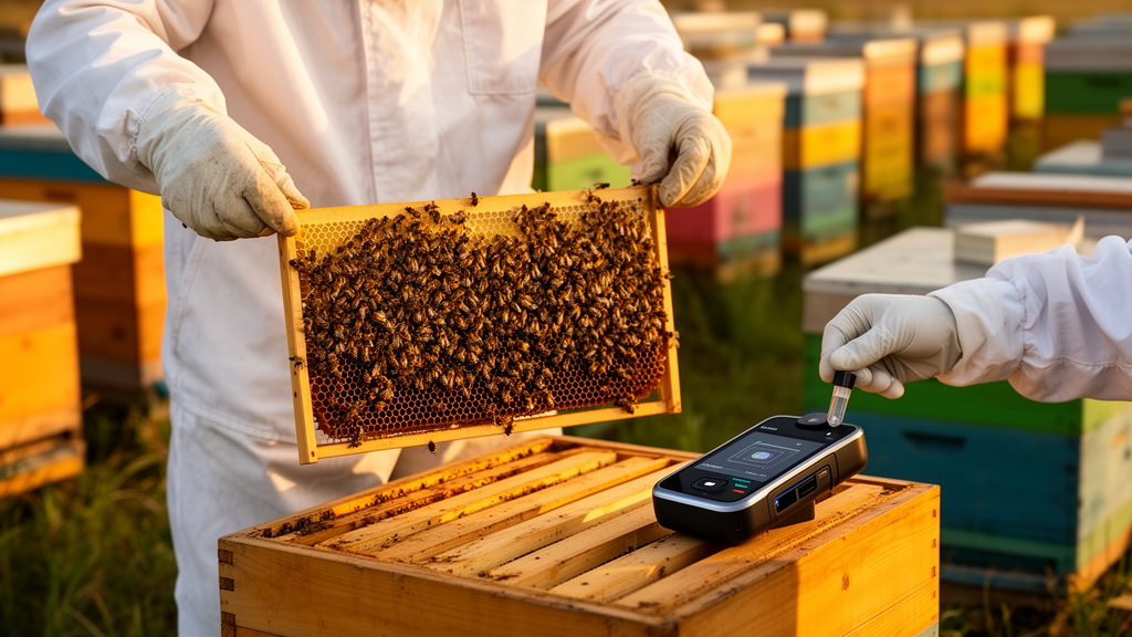
Inside a beehive, trouble rarely announces itself. By the time a beekeeper spots a patchy brood pattern or sunken, perforated cell caps, the pathogens behind the damage may have been quietly building for weeks. A new review in Frontiers in Bee Science argues that the tools of modern genomics, particularly 16S rRNA gene amplicon sequencing, could change that calculus, giving veterinarians and beekeepers a way to identify bacterial brood diseases before colonies reach the point of collapse.
The review, authored by Joerg Mayer of the Department of Small Animal Medicine and Surgery at the University of Georgia’s College of Veterinary Medicine, positions honey bees as an emerging frontier in exotic pet medicine. Veterinarians are increasingly consulted about managed colonies, yet the diagnostic toolkit available to them has remained largely unchanged for decades: visual inspection, mite counts, alcohol washes, and a handful of field tests such as the Holst milk test, the matchstick test for American foulbrood, and lateral flow devices where they are available. These methods are inexpensive and accessible, but each targets a single threat and often detects infection only at an advanced stage.
The technical logic behind 16S rRNA amplicon sequencing is straightforward. Rather than trying to grow bacteria on laboratory media, the method reads the sequence of a conserved gene present in all bacteria, using that genetic barcode to identify which organisms are present and in what relative proportions. A companion approach targeting the internal transcribed spacer region performs the same service for fungi. Because the technique does not require viable organisms, it can detect fastidious pathogens that resist culture, and a single run can profile the entire bacterial and fungal community of a sample simultaneously.
That last point matters enormously for European foulbrood, a disease caused by Melissococcus plutonius. The bacterium is notoriously difficult to culture, demanding precise anaerobic conditions and specific sodium and potassium ratios, and it proliferates almost exclusively in the midgut of living larvae. Once a larva dies, M. plutonius declines rapidly while hardier secondary invaders such as Paenibacillus and Enterococcus species, which tolerate oxygen and grow readily on standard media, take over the sample. Culture-based diagnostics therefore risk recovering the opportunistic followers while missing the primary pathogen entirely.
The review illustrates the technology’s promise through four clinical cases, all of which began with beekeepers requesting antibiotic treatment for suspected European foulbrood. In the first case, a strong colony with no overt clinical problems was monitored longitudinally. A February sample from the brood nest contained no detectable M. plutonius. Three months later, follow-up sequencing revealed low levels of the bacterium, but because no clinical signs were present, antibiotic treatment was withheld. By August, after the nectar flow in Georgia, the hive displayed a characteristic shotgun brood pattern, with patchy cells resembling scattered shot holes, and resampling showed a marked increase in M. plutonius abundance.
That colony received a probiotic treatment, and a one-month follow-up sample indicated reduced detection of the pathogen, coinciding with visible hive improvement. The author is careful to note a caveat: European foulbrood symptoms are strongly seasonal and often resolve later in the season regardless of intervention, so controlled studies would be required to attribute the improvement specifically to the probiotic. Even so, the case demonstrates the core value of longitudinal sequencing, the ability to track bacterial abundance over time rather than relying on a single snapshot.
The second case shows the technology working in the opposite direction, ruling out a suspected bacterial disease. A routine inspection revealed an abnormal brood pattern resembling European foulbrood, but 16S rRNA sequencing detected no bacterial pathogens consistent with the disease. Subsequent viral screening identified sacbrood virus, and closer examination confirmed typical sacbrood larval features. The finding redirected the diagnosis away from antibiotic intervention entirely, although the colony ultimately succumbed to the infection. Because mite management data were unavailable, the author notes that parasitic mite syndrome should also have been considered in the differential diagnosis.
The remaining two cases document co-infections that would have been difficult to untangle by culture. In one hive with an abnormal brood pattern, sequencing detected M. plutonius alongside substantial quantities of Enterococcus faecalis, an interpretation the author describes as European foulbrood with associated secondary bacterial overgrowth rather than a primary E. faecalis infection. The brood abnormalities in that hive could also have been consistent with queen failure or laying-worker activity, underscoring how many alternative explanations must be weighed. That hive perished weeks after sampling. In the fourth case, sequencing identified Paenibacillus dendritiformis together with M. plutonius in a co-infection. The bacterium is a common environmental microbe previously reported in the United States in sources such as clay, but this appears to be the first documented detection in honey bee colonies or larvae, with prior literature limited to Switzerland. Probiotic powder treatment was followed by a marked reduction in both organisms on follow-up testing, and the colony survived without antibiotics.
The review is equally candid about limitations. 16S rRNA sequencing reports relative abundance rather than absolute pathogen load, and semi-quantitative estimates carry biases related to PCR amplification, DNA extraction efficiency, and total microbial biomass. Bacterial composition can vary substantially between individual larvae and can shift rapidly after larval death, making proper sample handling and cold-chain maintenance essential. As with any DNA-based method, the technique cannot distinguish viable from non-viable organisms and does not by itself establish pathogenicity. Practical constraints, including cost, turnaround times that can exceed rapid assays, and the need for bioinformatics expertise, currently limit routine diagnostic use.
Interpretation, the author stresses, must always be integrated with colony-level context: queen status, colony strength, Varroa mite counts and management history, seasonality, and clinical examination findings. Detection of secondary bacteria such as Enterococcus species should never be read as evidence of causation without additional supporting data. Within those boundaries, the technology offers real advantages, including turnaround times of roughly two to three days from sample receipt to reporting, which can be faster than culture and susceptibility testing for fastidious organisms, and a transport buffer that stabilizes the microbial community at the moment of sampling, eliminating the viability requirements that plague conventional culture. The approach also supports antimicrobial stewardship at a moment when multidrug-resistant bacteria linked to antibiotic residues in animal-derived food products are a growing public health concern, and many countries already prohibit antibiotic use in beehives for managing foulbrood diseases. The author’s conclusion is measured but clear: 16S rRNA gene amplicon sequencing is best viewed as a complementary diagnostic adjunct rather than a replacement for conventional methods, one whose future value depends on standardization, validation against gold-standard techniques, and clinically actionable interpretive frameworks that can translate raw sequence data into decisions at the hive.
Subject of Research: Use of 16S rRNA gene amplicon sequencing for diagnosing bacterial brood diseases in honey bee colonies
Article Title: Application of next-generation sequencing (NGS) for disease diagnosis in the beehive
Article References: Mayer, J. (2026). Application of next-generation sequencing (NGS) for disease diagnosis in the beehive. Frontiers in Bee Science, 4, Article 1862765. https://doi.org/10.3389/frbee.2026.1862765
Image Credits: AI Generated
DOI: 10.3389/frbee.2026.1862765
Keywords: honey bees, next-generation sequencing, 16S rRNA, European foulbrood, Melissococcus plutonius, brood disease, veterinary diagnostics, microbiome, antimicrobial stewardship, colony health, Paenibacillus dendritiformis, apiculture
Cite Scienmag News
Juliet Wilcox. (October 16, 2026). DNA Sequencing Steps Into the Beehive to Catch Hive Diseases Early. Scienmag. https://scienmag.com/dna-sequencing-steps-into-the-beehive-to-catch-hive-diseases-early/
Juliet Wilcox. "DNA Sequencing Steps Into the Beehive to Catch Hive Diseases Early." Scienmag, 16 October 2026, https://scienmag.com/dna-sequencing-steps-into-the-beehive-to-catch-hive-diseases-early/. Accessed 16 October 2026.
Juliet Wilcox. "DNA Sequencing Steps Into the Beehive to Catch Hive Diseases Early." Scienmag. October 16, 2026. https://scienmag.com/dna-sequencing-steps-into-the-beehive-to-catch-hive-diseases-early/


