


…
…
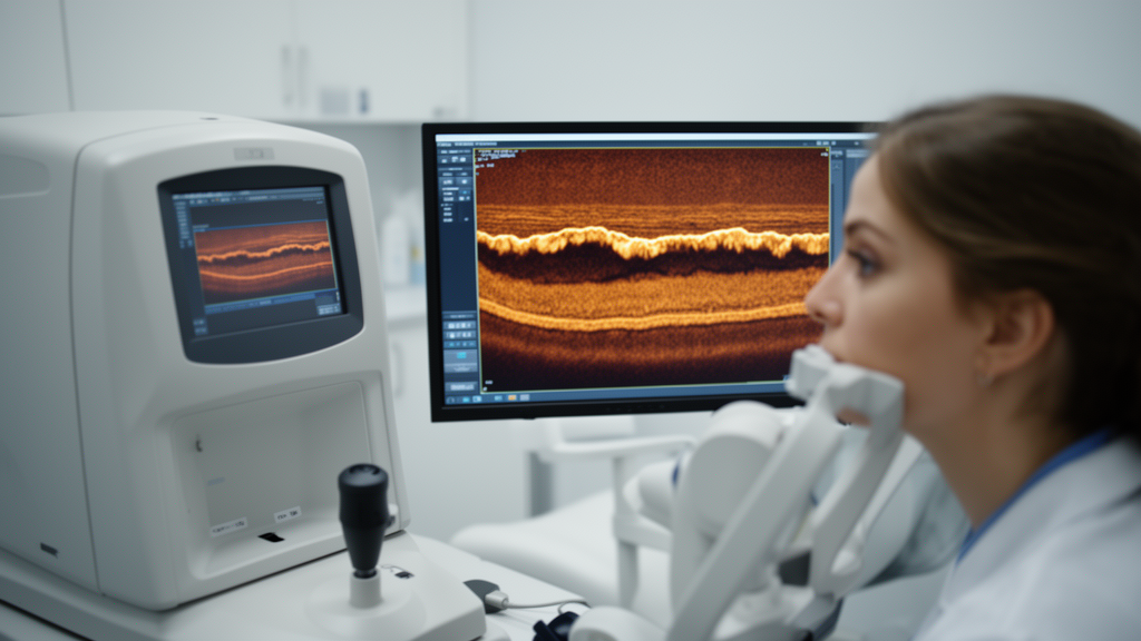
Optical coherence tomography has become one of the most widely used tools for studying the retina in systemic disease because it allows individual layers of tissue to be measured non-invasively and with micron-level precision. In the present study, the Spectralis platform resolved retinal structures at an axial resolution of 3.9 micrometers in tissue, which is fine enough to distinguish adjacent layers such as the ganglion cell layer and the inner plexiform layer that together measure only tens of micrometers in thickness. This level of resolution matters because diseases that affect the nervous system often produce their earliest detectable changes in specific layers rather than uniformly across the whole retina. The retinal nerve fiber layer, for example, consists of unmyelinated axons of retinal ganglion cells, and these axons share embryological, structural, and biochemical characteristics with central nervous system white matter. For this reason, the retina is frequently described as a window onto the brain, and layer-specific OCT measurements have been studied extensively in conditions such as multiple sclerosis, Parkinson disease, and Alzheimer disease, where thinning of the inner retinal layers is a common finding.
What makes the current findings noteworthy is the direction of the change. In many neurodegenerative conditions, OCT studies report thinning of the nerve fiber layer and ganglion cell complex, reflecting loss of neurons and their axons. In patients with Steinert disease, the study found a generalized thickening of retinal layers instead, together with significant thickening of the retinal nerve fiber layer in the temporal region of the optic nerve and in the papillomacular bundle. Thickening rather than thinning suggests that the pathological process in the retina of these patients may not be straightforward degeneration. Several mechanisms could theoretically produce increased retinal thickness, including intraretinal edema, accumulation of metabolic byproducts, altered cellular metabolism, gliosis involving Müller cells or astrocytes, or changes in axonal transport. Myotonic dystrophy type 1 is caused by toxic gain-of-function RNA from the expanded CTG repeat, which sequesters RNA-binding proteins and disrupts alternative splicing of many target genes. Because those splicing factors are active in multiple tissues, including neural and retinal tissue, the same molecular mechanism that produces myotonia in skeletal muscle could plausibly disturb cellular homeostasis in the retina, and the observed structural changes may reflect that disturbance rather than simple cell loss.
The papillomacular bundle deserves particular attention. This bundle contains the ganglion cell axons that run from the macula, the region responsible for central and high-acuity vision, to the optic disc, and it is known to be selectively vulnerable in several neurological disorders, including mitochondrial disease and dominant optic atrophy. The finding of significant RNFL thickening specifically in this bundle, detected with the aid of fovea-to-disc technology that aligns the scan to each patient’s individual anatomy, suggests a patterned rather than random involvement of the retinal nerve fiber layer. Patterned involvement is generally more informative than diffuse change because it can point to the underlying distribution of pathology. If future studies confirm that the papillomacular bundle is consistently affected in Steinert disease, this sector could serve as a region of interest in longitudinal monitoring, in the same way that specific peripapillary sectors are used to track glaucomatous progression.
The methodological choices in this study strengthen the reliability of its conclusions. Selecting only one eye per participant is an important safeguard, because the two eyes of a single individual are highly correlated in both anatomy and disease expression, and treating them as independent observations can artificially inflate statistical significance. Excluding eyes with axial lengths outside the range of 22.1 to 25.2 millimeters and refractive errors of five diopters or more of spherical equivalent controls for a well-known confounder, since axial elongation stretches the retina and thins many of its layers independently of any disease process. Restricting inclusion to patients with minimal lens opacity, scored 0 or 1 on the Lens Opacities Classification System III, reduces the risk that cataract-related signal degradation would distort the OCT segmentation. This last point is particularly relevant to Steinert disease, because cataracts are present in nearly all patients with the condition and could otherwise contaminate retinal measurements. Perhaps most importantly, the exclusion of eyes with epiretinal membranes and macular edema distinguishes this study from much of the earlier literature, in which such concomitant pathologies were often present and may have driven the retinal changes attributed to the disease itself.
The two imaging protocols used here complement each other in a useful way. The posterior pole protocol divides a 25 by 30 degree area of the macula into an 8 by 8 grid of 64 cells, each approximately one square millimeter, and segments eight distinct layers in every cell. This dense sampling allows localized changes to be detected that would be averaged away in a single global measurement. The Anatomic Positioning System corrects for head tilt and cyclotorsion of the eye by referencing a horizontal line between the fovea and the opening of Bruch’s membrane, ensuring that the grid is aligned consistently across patients and visits. The Nsite analytics module, in contrast, focuses on the peripapillary nerve fiber layer and divides it into six sectors, using the same fovea-to-disc alignment to standardize the position of the papillomacular bundle analysis. Together, these protocols cover both the macular and peripapillary territories, which is valuable because different diseases affect these regions differently; for example, ganglion cell involvement often shows first in the macula, while axonal changes may appear earlier around the disc.
The sample sizes, 79 eyes from 79 patients with genetically confirmed disease and 128 control eyes for the posterior pole analysis, are respectable for a study of a relatively rare condition. Myotonic dystrophy type 1 is the most common muscular dystrophy affecting adults, with an estimated prevalence in the range of one in several thousand in European populations, yet recruitment remains challenging because affected individuals may have limited mobility and because the clinical presentation varies widely with the size of the CTG repeat expansion. Larger repeat expansions are generally associated with earlier onset and more severe disease, a phenomenon known as anticipation, in which the repeat length can increase across generations. This heterogeneity means that future studies correlating retinal measurements with repeat length, age at onset, or the severity of muscle weakness and myotonia would need to account for these variables explicitly, and the present cross-sectional design was not powered to establish such correlations.
The authors are appropriately cautious on this point. No conclusive evidence currently links the retinal findings to the severity of neuromuscular symptoms, and the study does not claim that OCT measurements can substitute for neurological assessment. What the results do establish is that retinal structural alterations are present in patients with Steinert disease even when confounding ocular pathologies are excluded, and that these alterations are detectable with standard commercial OCT protocols. This opens several lines of future investigation. Longitudinal studies could determine whether retinal layer thickness changes over time and whether the rate of change relates to disease progression elsewhere. Correlation studies could test whether retinal measures track with markers of central nervous system involvement, such as cognitive testing or neuromuscular scores. The observation that the retina may reflect involvement of the broader nervous system is consistent with the multisystemic character of myotonic dystrophy, in which the central nervous system is affected in many patients, producing features such as cognitive impairment, excessive daytime sleepiness, and apathy.
From a clinical standpoint, the findings support the inclusion of routine ophthalmological evaluation in the care of patients with Steinert disease, beyond the assessment of cataracts that is already standard practice. Patients with this condition are known to have ptosis, weakness of the extraocular muscles, slow pupillary responses, and low intraocular pressure in addition to cataracts, and the current results add retinal structural changes to this list. Because the changes were detected in patients whose lenses were nearly clear, they are unlikely to be artifacts of media opacity, and because the control group was age-matched, they are unlikely to reflect normal aging. The color maps generated by the posterior pole protocol can visualize differences of 10 to 15 micrometers, which means that even subtle changes could be followed in clinical practice if validated thresholds and progression criteria are established.
It is worth noting some limitations inherent to the study design. The cross-sectional nature of the data means that the direction of causality between the disease and the retinal changes cannot be established, although the genetic basis of Steinert disease makes the direction reasonably clear in this case. The number of control eyes available for the peripapillary Nsite analysis was smaller, 39 eyes, than for the posterior pole analysis, because the additional normative database controls lacked Nsite recordings, and this asymmetry should be kept in mind when weighing the peripapillary findings. No manual correction of the automated segmentation was applied, which preserves objectivity but means that segmentation errors, if any occurred, were not corrected; the use of an internal fixation target, which has the highest reported reproducibility, helps to minimize such errors by stabilizing fixation during acquisition. Finally, the patients were recruited at a single center in Spain, and replication in independent cohorts with different ancestries and disease durations would strengthen the generalizability of the conclusions.
In summary, this study contributes a carefully controlled characterization of retinal layer structure in type 1 myotonic dystrophy, demonstrating generalized retinal thickening and sector-specific nerve fiber layer changes in the temporal optic nerve and papillomacular bundle. The work illustrates how high-resolution, layer-specific imaging can reveal patterns of involvement in a multisystemic genetic disease and provides a foundation for the use of OCT-derived structural markers in future research on retinal and possibly central nervous system involvement in Steinert disease.
Subject of Research: Retinal layer structural changes in Steinert disease measured by optical coherence tomography
Article Title: Structural changes in the retinal layers of patients with Steinert disease
Article References: Structural changes in the retinal layers of patients with Steinert disease. (n.d.). https://doi.org/10.3389/fopht.2026.1959492
Image Credits: AI Generated
DOI: 10.3389/fopht.2026.1959492
Keywords: Steinert disease, myotonic dystrophy type 1, optical coherence tomography, retinal layers, retinal nerve fiber layer, papillomacular bundle, neuro-ophthalmology, retina, biomarkers, DMPK gene, Structural, changes
Cite Scienmag News
Diana Fleming. (October 16, 2026). Optical coherence tomography reveals retinal layer thickening in Steinert disease patients. Scienmag. https://scienmag.com/optical-coherence-tomography-reveals-retinal-layer-thickening-in-steinert-disease-patients/
Diana Fleming. "Optical coherence tomography reveals retinal layer thickening in Steinert disease patients." Scienmag, 16 October 2026, https://scienmag.com/optical-coherence-tomography-reveals-retinal-layer-thickening-in-steinert-disease-patients/. Accessed 16 October 2026.
Diana Fleming. "Optical coherence tomography reveals retinal layer thickening in Steinert disease patients." Scienmag. October 16, 2026. https://scienmag.com/optical-coherence-tomography-reveals-retinal-layer-thickening-in-steinert-disease-patients/


