


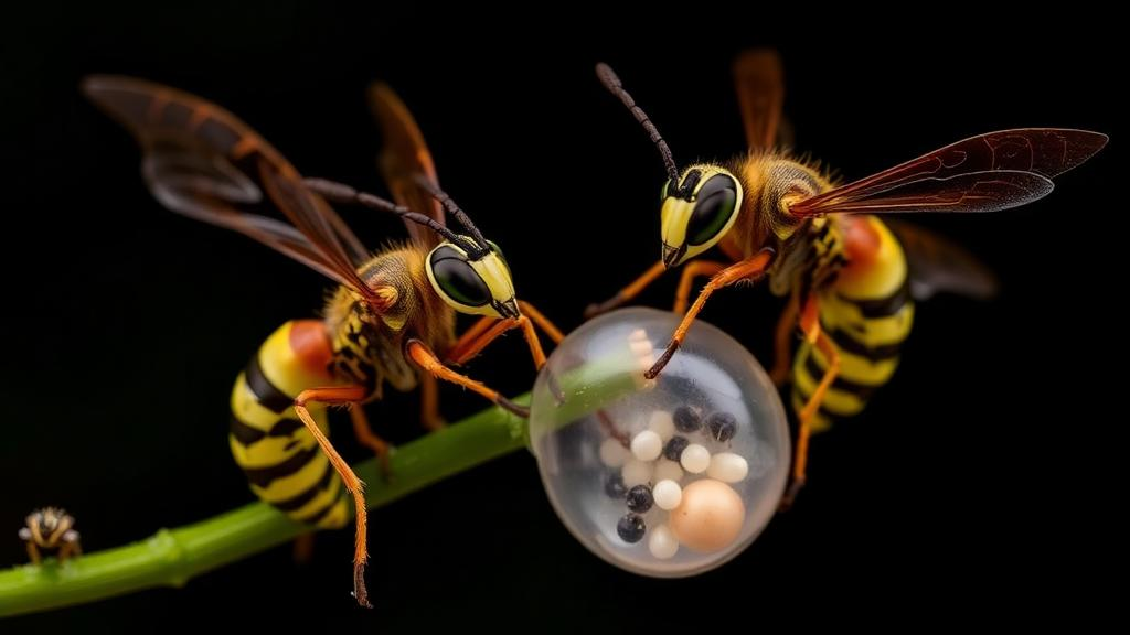
Tiny parasitic wasps are quietly rewriting one of biology’s oldest assumptions: that the earliest steps of an embryo’s life are too fundamental to change. A new study of the whitefly parasitoid Encarsia lutea, published in Frontiers in Insect Science, shows that even closely related species within a single genus can build their embryos in radically different ways. The findings, from a team of Italian researchers led by Francesco Nugnes and Paolo Pedata of the National Research Council and Antonio Garonna and Donato Mancini of the University of Naples Federico II, reveal a level of developmental diversity within one genus that, for most of the insect order they belong to, is normally seen only when comparing entire families.
The researchers set out to chart the complete embryogenesis of Encarsia lutea, a solitary, arrhenotokous endoparasitoid that attacks economically devastating whiteflies such as the greenhouse whitefly Trialeurodes vaporariorum and the silverleaf whitefly Bemisia tabaci. Using light and fluorescence microscopy on embryos collected at precise intervals from five minutes to ninety hours after the host was stung, and staining both F-actin filaments, which mark cellular boundaries, and nuclei with fluorescent dyes, they reconstructed every major event from fertilization to the moment the membrane around the larva dissolves. The result is one of the most detailed cell-by-cell accounts of early development ever compiled for the chalcid wasps, a group whose embryology remains poorly known outside a handful of species.
What they found was a creature caught evolutionarily in the middle. Insects typically build their eggs the same way: yolk-protein precursors made by the mother are packed into a central mass, the embryo’s nuclei divide and migrate to the egg’s periphery, and development proceeds with minor variations on the Drosophila theme. E. lutea breaks that mold only partway. Its freshly laid egg measures just 88.3 by 23.7 micrometers and is oligolecithal, meaning it carries a reduced, though not absent, supply of yolk. Within minutes of deposition, the egg begins swelling, drinking fluid from the host’s body cavity in a process called hydropic behavior. It occupies a genuine intermediate position: the egg of its congener Encarsia formosa is fully yolk-rich and does not swell, while that of Encarsia pergandiella carries essentially no yolk at all and expands dramatically after oviposition.
Early events in E. lutea followed the canonical script closely. The sperm entered the egg within the first hour and lost its tail, the female’s meiotic divisions produced the first and second polar bodies, and the male and female pronuclei fused into a synkaryon between the second and third hour. The first two cleavage nuclei appeared between the third and fourth hours, and thereafter the embryo divided in the peripheral pattern typical of insects with yolk-laden eggs, with most nuclei migrating toward the surface to form a blastoderm. But then came a surprise. At the eight-nucleus stage, one nucleus sitting at the apex of the embryo was walled off inside its own membrane by contraction of an actin cortex, creating a separate apical blastomere. Its four daughter nuclei remained sharply segregated from the rest of the embryo throughout subsequent development.
The apical blastomere was only the first act of a much larger drama. Around the twenty-fourth hour, cells in the ventral region of the blastoderm elongated into a columnar shape and, within hours, gave rise to a layer of some fifty to sixty unusually large cells bearing giant nuclei, a signature of polyploidy, in which the genome is replicated repeatedly without cell division. These large cells spread beneath the chorion and, together with the descendants of the apical blastomere, eventually wrapped the developing larva in a continuous extra-embryonic membrane, or EEM. Such membranes are hallmarks of endoparasitic wasps: they protect the embryo from the host’s immune system, regulate host physiology, and deliver nutrients from the host’s blood.
At hatching, around ninety hours after oviposition, the membrane performed its final trick. It dissociated into fifty to sixty teratocytes, uniform cells with a mean diameter of 22.8 micrometers that float freely in the host’s hemolymph. Teratocytes are among the most remarkable weapons in a parasitoid’s arsenal. They secrete proteins that suppress the host’s immune defenses, manipulate its hormones, and provide nutrition to the growing larva; a proteotranscriptomic study of another species found that teratocytes can release dozens of distinct polypeptides into the host. In E. lutea, the teratocyte population then behaved in the classic manner: their numbers fell steadily, from roughly fifty-six at hatching to about thirty-five in the second larval stage and fewer than thirty in the mature larva, while their average size grew from about twenty-three to nearly thirty-eight micrometers, consistent with cells being lost through larval ingestion, degeneration, or host expulsion rather than division.
The true significance of these observations emerges only in comparison with the two other Encarsia species whose embryology has been studied in comparable depth, and here the picture becomes genuinely striking. All three species share meroistic polytrophic ovaries, the standard setup for holometabolous insects, yet they differ in how much yolk each egg carries and, consequently, in almost every downstream developmental event. E. formosa, with a yolk-rich anhydropic egg, develops through the textbook peripheral cleavage with no extra-embryonic membrane and no teratocytes. E. pergandiella, with its yolkless egg, undergoes holoblastic cleavage, the complete subdivision of the entire egg, and assembles its membrane from several cleavage nuclei, including derivatives of the polar bodies, producing only about ten enormous teratocytes ranging from fifty-five to eighty-five micrometers. E. lutea borrows features from both but follows its own path: peripheral cleavage like E. formosa, hydropic swelling and a membrane with teratocytes like E. pergandiella, yet a membrane whose majority lineage arises not from early-cleavage nuclei but from a later differentiation event within the blastoderm itself.
That difference in origin matters for how biologists classify these structures. In parasitic Hymenoptera, extra-embryonic membranes have been sorted into categories defined by their embryonic source: the serosa from extraembryonic blastoderm cells, the pseudoserosa from delamination of an undifferentiated blastoderm, the deutoserosa from yolk-associated internal cells, and the trophamnion from polar body derivatives. The authors suggest that the blastoderm-derived portion of the E. lutea membrane, formed through a process resembling simultaneous delamination followed by a switch from mitotic division to endoreplication, fits the pseudoserosa. But the apical portion, descended from that early segregated blastomere, matches none of the established categories cleanly. They also note, intriguingly, that in both E. lutea and E. pergandiella the polar bodies appear anchored by actin cortex at the apical border precisely where the early blastomere segregates, raising the possibility that the polar bodies themselves help specify embryonic cell fate, a role recently demonstrated in mammals.
The broader lesson is that early embryogenesis in parasitoid wasps is not phylogenetically locked in place. The shift from external to internal parasitism has long been proposed as the main engine of these changes, because an embryo floating in a host’s blood can abandon the yolk its ancestors needed and absorb nutrients directly. Loss of the vitellogenin gene, which encodes the yolk protein precursor, has occurred independently in at least five unrelated parasitoid lineages. Yet the picture is not simple: recent genomic work found that about thirty-five percent of endoparasitoids retain vitellogenin copies, and within Encarsia itself, yolk-rich eggs remain widespread among the genus’s 486 described species. The authors favor a more nuanced view in which phylogenetic history, environmental interactions, internal constraints, and genetic flexibility jointly determine which developmental features are conserved and which are free to change. They also flag the possibility of developmental system drift, the phenomenon in which underlying genetic and cellular mechanisms diverge dramatically even as the final adult form stays essentially unchanged.
Perhaps the most provocative implication is temporal. Comparative evidence suggests that some of the developmental strategies seen in Encarsia species represent transitional conditions, snapshots of evolution in progress toward the complete loss of yolk production, with E. lutea standing partway between the two extremes its relatives have already reached. The authors even point out that holoblastic cleavage, seen in the most derived endoparasitoids, resembles the pattern of the most basal hexapods, hinting at possible back-and-forth transitions between total and superficial cleavage across arthropod evolution. With the raw data now publicly available and modern tools of sequencing, transcriptomics, and gene silencing increasingly applicable to these tiny wasps, the authors argue that the megadiverse genus Encarsia is ideally positioned as a microevolutionary model, one small enough to compare sibling species directly yet diverse enough to test the biggest questions about how natural selection reshapes the very first hours of life.
Subject of Research: Embryonic development and extra-embryonic membrane formation in the parasitoid wasp Encarsia lutea compared with congeners
Article Title: Building the parasitic wasp Encarsia: a tale of three tails
Article References: Building the parasitic wasp Encarsia: a tale of three tails. (n.d.). https://doi.org/10.3389/finsc.2026.1861915
Image Credits: AI Generated
DOI: 10.3389/finsc.2026.1861915
Keywords: Encarsia lutea, parasitoid wasps, embryogenesis, extra-embryonic membranes, teratocytes, cleavage patterns, hydropic eggs, vitellogenin, Hymenoptera, evolution of development, whitefly parasitoid, EvoDevo
Cite Scienmag News
Gavin Prescott. (October 15, 2026). Three Sister Wasp Species, Three Wildly Different Ways to Build an Embryo. Scienmag. https://scienmag.com/three-sister-wasp-species-three-wildly-different-ways-to-build-an-embryo/
Gavin Prescott. "Three Sister Wasp Species, Three Wildly Different Ways to Build an Embryo." Scienmag, 15 October 2026, https://scienmag.com/three-sister-wasp-species-three-wildly-different-ways-to-build-an-embryo/. Accessed 15 October 2026.
Gavin Prescott. "Three Sister Wasp Species, Three Wildly Different Ways to Build an Embryo." Scienmag. October 15, 2026. https://scienmag.com/three-sister-wasp-species-three-wildly-different-ways-to-build-an-embryo/


