


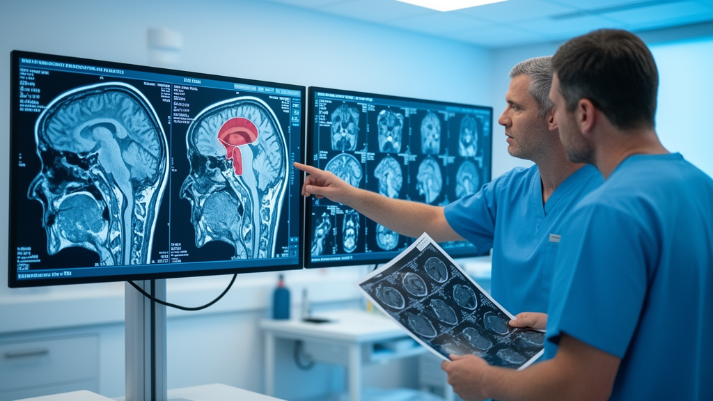
Deep in the brainstem, wrapped around the very fibers that let us walk, reach, and grip, sit some of the most dangerous tumors in neurosurgery. Brainstem gliomas are rare, but their location makes every operation a high-stakes gamble: surgeons must remove cancerous tissue while tiptoeing around the corticospinal tract, the bundle of nerve fibers carrying the brain’s motor commands down to the muscles. Now a team at Beijing Tiantan Hospital, Capital Medical University, has shown that a preoperative MRI technique can sort these patients into groups with dramatically different recoveries, offering surgeons a way to predict, before the first incision, whose motor function will bounce back and whose will not.
The study, published in the Journal of Neuro-Oncology, followed 180 patients who underwent craniotomy for brainstem glioma between 2020 and 2023. Before surgery, each patient received diffusion tensor imaging, or DTI, a specialized MRI method that tracks the direction water molecules diffuse through brain tissue. Because water moves more freely along the length of a nerve fiber than across it, DTI can reconstruct the three-dimensional course of white matter tracts, a process called tractography. The researchers used this technique to classify how each tumor interacted with the corticospinal tract, sorting the cases into three categories: displacement, infiltration, and traverse.
The classification itself is elegantly simple. In the displacement type, the tumor pushes the corticospinal tract aside without breaching it, like a growing root displacing a buried pipe. In the infiltration type, tumor cells invade the tract’s territory, blurring the boundary between cancer and conduit. In the traverse type, the tumor cuts straight through the tract, splitting the motor highway in two. Two independent raters applied the scheme with near-perfect agreement, achieving an inter-rater reliability coefficient of 0.93, a level of consistency that suggests the system could be reproduced reliably in hospitals anywhere.
The technical measurements behind the categories revealed stark biological gradients. Fractional anisotropy, a DTI metric that quantifies how directional water diffusion is within a tract, served as a proxy for the structural integrity of the corticospinal tract. Displacement patients showed the highest values, averaging 0.45, while traverse patients had the lowest, at 0.424, reflecting tracts already damaged by the tumor cutting through them. Infiltration patients fell in between. These differences were not subtle statistical noise; the gradients across subtypes were highly significant, and fractional anisotropy correlated positively with preoperative muscle strength measured on the Medical Research Council scale.
Tumor biology tracked the imaging categories with remarkable fidelity. Among traverse-type patients, 97.8 percent carried high-grade tumors, the aggressive, fast-growing kind. Among displacement patients, 74.5 percent had low-grade tumors, the slower-growing variety. Infiltration occupied the middle ground. Preoperative functional status followed the same pattern: displacement patients arrived at the hospital with the best Karnofsky Performance Status and modified Rankin Scale scores, meaning they were largely independent in daily life, while traverse patients were the most disabled before surgery ever began.
What sets this study apart from earlier imaging work is its longitudinal design. Rather than capturing a single snapshot, the team collected functional data at predefined intervals after surgery, using the Karnofsky Performance Status, the modified Rankin Scale, and MRC muscle strength grades, and analyzed the recovery curves with linear mixed-effects models. This statistical approach, which handles repeated measurements from the same patients over time, revealed that the three groups did not merely start at different points; they followed fundamentally different trajectories through recovery, a finding that held up after correction for multiple comparisons.
The trajectories tell three very different stories. Displacement patients recovered rapidly, regaining motor function at a pace consistent with a tract that was structurally intact but merely pushed aside. Traverse patients showed minimal recovery and, strikingly, experienced late decline in function, a pattern consistent with progressive tract degeneration rather than healing. Infiltration patients traced an intermediate course. For the first time, surgeons can look at a preoperative scan, assign a category, and have a data-driven expectation of how the months after surgery will unfold for that patient’s ability to move.
Survival data reinforced the imaging-based stratification. Median overall survival was not reached in the displacement group, meaning most of these patients were still alive when the analysis was performed. Infiltration patients survived a median of 15.3 months, and traverse patients just 12.0 months. All pairwise comparisons between the groups were statistically significant. The finding echoes a broader principle in neuro-oncology: surgically acquired motor deficits are associated with worse survival, likely because functional decline limits access to adjuvant therapies and erodes quality of life in the time that remains.
The implications reach beyond the operating room. A preoperative classification that integrates tumor-tract interaction could reshape surgical planning, from choosing safe entry zones into the brainstem to deciding how aggressively to pursue resection. For displacement-type patients, whose tracts are intact and whose tumors are often low-grade, the data support pursuing maximal safe removal with confidence in functional recovery. For traverse-type patients, whose tracts are already breached and whose tumors are overwhelmingly high-grade, the framework raises the question of whether aggressive surgery serves the patient, or whether biopsy, radiation, and palliative pathways deserve earlier consideration. The authors describe the system as hypothesis-generating, a careful phrase that acknowledges the study was observational and that prospective validation is needed before the classification becomes standard practice.
Still, the study represents a meaningful step toward personalized neurosurgery for one of medicine’s most forbidding tumor locations. Brainstem gliomas have long resisted the progress seen elsewhere in glioma care, partly because the brainstem’s dense functional anatomy makes both surgery and imaging interpretation difficult. By turning the corticospinal tract from a hazard to be avoided into a measurable prognostic variable, the Beijing team has given clinicians a framework grounded in physics, statistics, and 180 carefully followed patients. If future studies validate these recovery trajectories, the humble DTI scan, already standard in most hospitals, could become the first tool that tells a brainstem glioma patient, before surgery, what their road to recovery will look like.
Subject of Research: Longitudinal motor recovery trajectories in brainstem glioma associated with preoperative DTI-based corticospinal tract involvement patterns
Article Title: Longitudinal motor recovery trajectories in brainstem glioma associated with preoperative DTI-based corticospinal tract involvement patterns
Article References: Longitudinal motor recovery trajectories in brainstem glioma associated with preoperative DTI-based corticospinal tract involvement patterns. (n.d.). https://doi.org/10.1007/s11060-026-05829-4
Image Credits: AI Generated
DOI: 10.1007/s11060-026-05829-4
Keywords: brainstem glioma, diffusion tensor imaging, corticospinal tract, neurosurgery, motor recovery, prognosis, brain cancer, Longitudinal, motor, recovery, trajectories, brainstem
Cite Scienmag News
Nathaniel Bowman. (October 15, 2026). Brainstem Tumor Scans Predict Which Patients Will Walk Again After Surgery. Scienmag. https://scienmag.com/brainstem-tumor-scans-predict-which-patients-will-walk-again-after-surgery/
Nathaniel Bowman. "Brainstem Tumor Scans Predict Which Patients Will Walk Again After Surgery." Scienmag, 15 October 2026, https://scienmag.com/brainstem-tumor-scans-predict-which-patients-will-walk-again-after-surgery/. Accessed 15 October 2026.
Nathaniel Bowman. "Brainstem Tumor Scans Predict Which Patients Will Walk Again After Surgery." Scienmag. October 15, 2026. https://scienmag.com/brainstem-tumor-scans-predict-which-patients-will-walk-again-after-surgery/


