


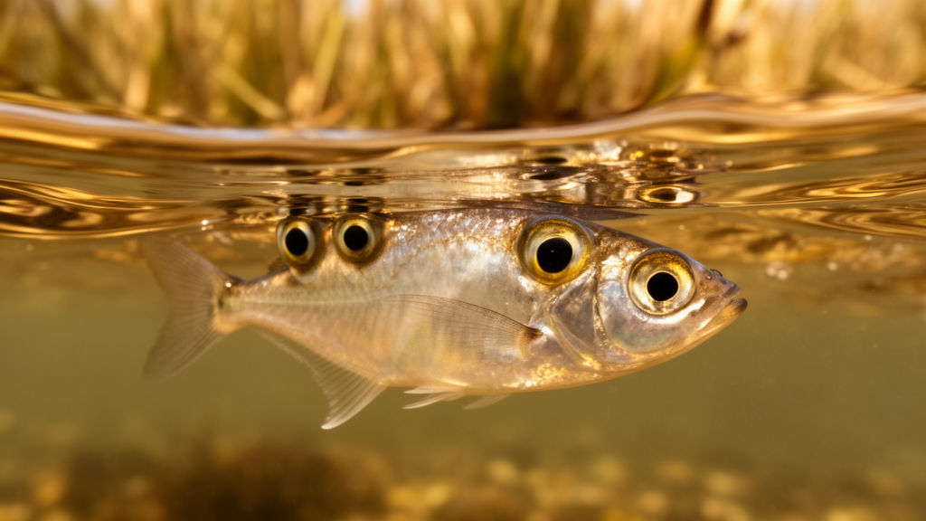
Along the muddy margins of rivers and mangrove channels in Central and South America swims one of the strangest eyes in the vertebrate world. The four-eyed fish, Anableps anableps, does not actually have four eyes, but each of its two eyes is divided into two functionally separate halves by a band of tissue, allowing the fish to simultaneously survey the world above the waterline and the world beneath it. As the fish cruises at the surface with the top of its eye protruding into the air, the lower half of each retina receives light that has traveled through water, while the upper half of the retina, oriented downward in the eye’s cup, processes light arriving from the air. Now, a team of researchers at Louisiana State University has traced how this remarkable dual-vision system evolved at the level of genes and developing tissue, revealing that Anableps achieved its aerial view not by inventing new cell types, but by expanding and reprogramming a region of the retina it already had.
The study, published in Nature Communications by Louise Perez, Lina Shi, and colleagues in the laboratory of Patricia N. Schneider, tackles one of the deepest questions in biology: how complex organs acquire genuinely new capabilities during evolution. The vertebrate eye is often cited as an example of intricate engineering, with its layered neurons, lens, and light-sensitive photoreceptors, and understanding how such a structure can be modified without breaking it is a central challenge for evolutionary developmental biology. Anableps offers an ideal natural experiment, because the species lives its life straddling two optical environments at once. Light behaves very differently in air and in water, bending to different degrees as it enters the eye and carrying different spectral information, so a retina built for underwater vision is not automatically well suited to aerial vision.
To find out what changed in the Anableps eye, the researchers turned to two of the most powerful tools in modern genomics: single-nucleus transcriptomics and spatial transcriptomics. Single-nucleus methods allow scientists to catalog which genes are switched on in thousands of individual cells, revealing the full diversity of cell types present in a tissue. Spatial transcriptomics goes a step further by mapping where those gene-expression profiles sit within the intact organ, preserving the anatomical geography that conventional sequencing destroys. Applied to the Anableps retina, these approaches produced a cell-by-cell, position-by-position atlas of gene activity, letting the team compare the dorsal half of the retina, which handles underwater light, with the ventral half, which handles aerial light.
The atlas delivered a striking first finding: the ventral retina, the region devoted to the aerial view, is developmentally expanded. In a conventional fish eye, the dorsal and ventral halves of the retina are patterned by conserved developmental signals that establish the organ’s top-bottom axis during embryogenesis. In Anableps, the team found that markers of ventral retinal identity are overrepresented, and comparative spatial profiling of developing and adult Anableps alongside the killifish, a related surface-dwelling species with an ordinary single-vision eye, showed that the boundary separating the dorsal and ventral territories has shifted toward the dorsal side. The result is a proportionally larger ventral domain. In other words, evolution did not build a new compartment in the eye; it redrew the existing map, enlarging the aerial-vision territory at the expense of the aquatic one.
The second major finding concerns the photoreceptors themselves, the cone cells that provide color vision in bright light. Cones owe their spectral sensitivity to light-absorbing proteins called opsins, and the wavelength each opsin absorbs best is determined by its amino acid sequence. Fish genomes typically harbor multiple opsin genes, or paralogs, that arose through ancient duplications, and different copies can be tuned to different parts of the light spectrum. The spatial transcriptomic data revealed that opsin expression in Anableps is regionally organized within a single cone subtype. Cones in the ventral, aerial-vision region of the retina, designated PR1 cones, express a gene called opn1mw2, whereas the corresponding cones in the dorsal, underwater region express the longer-wavelength-sensitive opn1lw paralogs.
That regional swap matters because the two optical environments demand different sensitivities. To estimate how the proteins actually perform, the researchers used machine-learning predictions of opsin spectral properties. These analyses indicated that the Opn1mw2 pigment found in the ventral cones is red-shifted relative to Opn1mw1, another member of the medium-wavelength-sensitive opsin family, but remains blue-shifted relative to the dorsal Opn1lw pigments. The ventral retina therefore occupies an intermediate spectral position: tuned differently from the dorsal retina, and presumably better matched to the quality of light arriving from above the surface, yet still drawing on the same family of cone pigments that ancestral fishes used. The adaptation is a reshuffling of existing genetic resources, not the appearance of an entirely new photoreceptor class.
Taken together, the results sketch a coherent evolutionary pathway. Anableps did not need to evolve novel genes or novel cell types to peer into the air. Instead, it co-opted the developmental machinery that already patterns the dorsal-ventral axis of the vertebrate retina, shifting the boundary between territories so that the ventral domain grew larger, and then rewired the opsin expression programs of conserved cone subtypes so that each half of the retina is tuned to its own light environment. This kind of remodeling, in which ancient developmental circuits are redeployed to produce new proportions and new regional specializations, is increasingly recognized as a common route to evolutionary novelty, particularly in organs as complex and constrained as the eye.
The study also highlights how non-model organisms can illuminate questions that laboratory staples cannot. Anableps is not a traditional genetic model like the zebrafish or the mouse, but its unusual lifestyle makes it uniquely informative about sensory adaptation. By pairing modern transcriptomic techniques with careful comparisons against a related fish with conventional vision, the researchers could distinguish what is genuinely unusual about the Anableps retina from what is shared across teleosts. The finding that dorsal-ventral patterning markers reveal an expanded ventral identity, corroborated by the spatial comparison with killifish at both developmental and adult stages, gives the conclusion a firm anatomical footing rather than resting on gene-expression differences alone.
For the four-eyed fish itself, the payoff is a life lived at the interface. Cruising the surface with half of each eye in each world, Anableps can scan for insects and predators in the air while simultaneously watching for threats and prey below, a capability that depends on a retina partitioned into two spectrally and developmentally distinct zones. The new work shows that this partition is written into the organ’s developmental blueprint and into the pigment choices of its cone cells. It is a vivid reminder that evolution is often a tinkerer rather than an inventor, achieving dramatic functional change by enlarging, shifting, and reprogramming the parts it already has.
The research, funded by start-up funds from Louisiana State University and a grant from the National Science Foundation’s Integrative Organismal Systems program, was published as an open-access article, making the full dataset and methods available to other teams studying retinal evolution and sensory ecology. As spatial and single-cell technologies continue to spread beyond traditional model species, eyes like those of Anableps are likely to yield further secrets about how vertebrate sensory organs adapt to the world’s many light environments, one developmental tweak at a time.
Subject of Research: Evolutionary remodeling of the ventral retina enabling simultaneous aerial and aquatic vision in the four-eyed fish Anableps anableps
Article Title: Evolutionary remodeling of the ventral retina enables aerial vision in the four-eyed fish Anableps anableps
Article References: Evolutionary remodeling of the ventral retina enables aerial vision in the four-eyed fish Anableps anableps. (n.d.). https://doi.org/10.1038/s41467-026-78775-4
Image Credits: AI Generated
DOI: 10.1038/s41467-026-78775-4
Keywords: four-eyed fish, Anableps anableps, retina, evolutionary developmental biology, opsins, spatial transcriptomics, single-nucleus transcriptomics, photoreceptors, aerial vision, dorsal-ventral patterning, non-model organisms, Nature Communications
Cite Scienmag News
Juliet Wilcox. (October 15, 2026). How the Four-Eyed Fish Rebuilt Its Retina to See in Air and Water at Once. Scienmag. https://scienmag.com/how-the-four-eyed-fish-rebuilt-its-retina-to-see-in-air-and-water-at-once/
Juliet Wilcox. "How the Four-Eyed Fish Rebuilt Its Retina to See in Air and Water at Once." Scienmag, 15 October 2026, https://scienmag.com/how-the-four-eyed-fish-rebuilt-its-retina-to-see-in-air-and-water-at-once/. Accessed 15 October 2026.
Juliet Wilcox. "How the Four-Eyed Fish Rebuilt Its Retina to See in Air and Water at Once." Scienmag. October 15, 2026. https://scienmag.com/how-the-four-eyed-fish-rebuilt-its-retina-to-see-in-air-and-water-at-once/


