


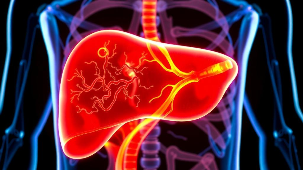
A new study from King’s College Hospital London in Dubai adds a compelling piece to one of the most debated puzzles in modern hepatology: whether vitamin D, the so-called sunshine hormone, is merely a bystander in fatty liver disease or an active participant in its progression toward irreversible scarring. The research, published in BMC Endocrine Disorders, examined 301 adult patients with metabolic dysfunction-associated steatotic liver disease, or MASLD, and found that nearly seven in ten were deficient in vitamin D. Those deficient patients were significantly more likely to have more severe fibrosis and more advanced fatty change in their livers, yet when the researchers carefully adjusted for confounding factors, vitamin D deficiency lost its status as an independent predictor of clinically significant scarring. The finding is a masterclass in the difference between association and causation, and it carries real implications for how clinicians think about a disease that now affects roughly a quarter to a third of all adults worldwide.
MASLD, formerly known under the umbrella of non-alcoholic fatty liver disease, has quietly become the most common chronic liver condition on the planet. It arises when fat accumulates inside liver cells in the context of metabolic problems such as obesity, insulin resistance, diabetes, and dyslipidaemia. In many people the fat deposition remains benign, but in a substantial subset it triggers inflammation, cell death, and the gradual activation of hepatic stellate cells, the resident cells responsible for producing scar tissue. That scarring, or fibrosis, is the single most important determinant of long-term outcomes: the more advanced the fibrosis, the higher the risk of cirrhosis, liver failure, and hepatocellular carcinoma. Identifying modifiable factors that accelerate or mark fibrosis progression is therefore one of the field’s most urgent priorities, and vitamin D has long been a candidate.
The biological rationale for suspecting vitamin D is genuinely intriguing. The vitamin D receptor is expressed on hepatic stellate cells, and experimental work has suggested that when activated vitamin D binds to this receptor, it can suppress the fibrogenic programs that turn quiescent stellate cells into collagen-producing myofibroblasts. Vitamin D also modulates immune responses and insulin signaling, both of which are central to the inflammatory milieu that drives MASLD progression. Deficiency is exceptionally common in people with metabolic disorders, partly because vitamin D is sequestered in adipose tissue, partly because sedentary indoor lifestyles reduce cutaneous synthesis, and partly because dietary intake is often inadequate. In Middle Eastern populations in particular, despite abundant sunshine, cultural and climatic factors conspire to produce some of the highest deficiency rates recorded anywhere in the world.
To interrogate the relationship rigorously, the research team turned to transient elastography, a non-invasive imaging technique marketed widely under the brand name FibroScan. The technology works by inducing gentle vibrations on the skin over the liver and measuring how fast shear waves propagate through the organ. Because stiff, scarred tissue transmits vibrations faster than healthy, fatty tissue, the resulting liver stiffness measurement provides a reliable surrogate for fibrosis stage, graded on a scale from F0, meaning no fibrosis, to F4, meaning cirrhosis. The same device estimates hepatic fat content through the controlled attenuation parameter, or CAP, which quantifies how strongly ultrasound waves are attenuated as they pass through the liver. Combining these two readouts allowed the investigators to assess both scarring and steatosis without subjecting patients to liver biopsy, the invasive gold standard that most large studies now seek to avoid.
The study population consisted of adults with MASLD who underwent vibration-controlled transient elastography and had a serum 25-hydroxyvitamin D measurement taken within ninety days, ensuring that the vitamin D status and the liver assessment reflected the same physiological moment. The cohort was relatively young, with a mean age of just under forty-two years, and nearly evenly split between sexes. The distribution of fibrosis was encouragingly skewed toward the mild end: roughly eighty-one percent of patients had no or mild fibrosis, about fifteen percent had significant fibrosis at stage F2, under four percent had advanced fibrosis at stage F3, and fewer than one percent had cirrhosis. Nevertheless, vitamin D deficiency was strikingly prevalent, affecting almost seventy percent of the entire cohort, a figure consistent with regional epidemiological data but still remarkable when viewed against the backdrop of a sun-drenched Gulf city.
When the researchers cross-tabulated vitamin D status against disease severity, the statistical signals were clear. Deficiency was significantly associated with fibrosis stage, with a p-value of 0.01, and independently associated with the severity of hepatic steatosis as quantified by CAP, also with a p-value of 0.01. Intriguingly, the associations with individual metabolic comorbidities were absent: body mass index, diabetes or prediabetes, hypertension, and dyslipidaemia showed no significant relationship with vitamin D deficiency in this cohort. What did stand out was metabolic syndrome as a composite diagnosis, which was strongly associated with deficiency at a p-value below 0.01. This pattern hints that vitamin D deficiency may track with overall metabolic burden rather than with any single component, a nuance that could matter for interpreting previous studies that focused on isolated risk factors.
The critical test came in the form of multivariable logistic regression, where vitamin D deficiency was evaluated as a predictor of clinically significant fibrosis while accounting for age, sex, and other covariates. The result was a resounding null: the adjusted odds ratio for vitamin D deficiency was 0.89, with a ninety-five percent confidence interval spanning 0.44 to 1.80 and a p-value of 0.754. In plain terms, once other factors were controlled for, knowing a patient’s vitamin D status added essentially no predictive value for identifying meaningful liver scarring. Instead, two far more conventional variables emerged as independent drivers. Each additional year of age increased the odds of significant fibrosis by about five percent, with an adjusted odds ratio of 1.05, and male sex carried a dramatically elevated risk, with an adjusted odds ratio of 6.23, meaning men in this cohort were more than six times as likely as women to have clinically significant fibrosis.
Why does this matter, and why should the finding still be considered news? The answer lies in the recalibration of expectations. Observational studies across many chronic diseases have linked low vitamin D to worse outcomes, fueling a wave of supplementation trials that have, almost uniformly, failed to deliver the benefits that the observational data promised. The present study adds liver fibrosis to the growing list of conditions where the crude association is real but likely non-causal. Vitamin D deficiency may function as a marker, a biological footprint of greater adiposity, reduced physical activity, more severe metabolic derangement, or simply older and sicker populations, rather than as an agent of harm in its own right. Treating it as such a marker could still be useful: a deficient patient with MASLD might warrant closer surveillance for fibrosis progression, even if correcting the deficiency itself is not expected to halt scarring.
The authors are careful and appropriately restrained in their conclusions. They explicitly state that causality cannot be established from this retrospective cross-sectional design, and they call for prospective studies to determine whether vitamin D plays any genuine role in disease progression. Such studies would need to follow patients over time, measure serial liver stiffness, and ideally randomize supplementation in deficient individuals to test whether repletion alters the fibrosis trajectory. Until then, the practical message for clinicians is nuanced: vitamin D deficiency is ubiquitous in MASLD patients, it correlates with disease severity on simple analysis, and it deserves correction for bone health and general wellbeing, but it should not be mistaken for a therapeutic lever against liver scarring. Meanwhile, the strongest signals in this data, age and male sex, serve as a reminder that the demographic profile of the patient in front of you may tell you more about their fibrosis risk than any single laboratory value.
For the millions of people living with MASLD worldwide, the study offers both reassurance and direction. The vast majority of patients in this cohort had no or only mild fibrosis, suggesting that early disease, when detected, remains the norm and the window for intervention is wide. The research also validates transient elastography as a practical, biopsy-free tool for stratifying risk in routine clinical settings, particularly in regions where deficiency and fatty liver overlap so extensively that disentangling their contributions demands precisely this kind of quantitative, mechanistically informed approach. The sunshine vitamin, it turns out, may simply be reflecting the light of underlying metabolic disease rather than driving the darkness of liver scarring, and knowing the difference is the first step toward treating the right target.
Subject of Research: Association between vitamin D deficiency and liver fibrosis in metabolic dysfunction-associated steatotic liver disease assessed by transient elastography
Article Title: Association between vitamin D deficiency and liver fibrosis assessed by transient elastography in patients with metabolic dysfunction-associated steatotic liver disease
Article References: Jose, J., Muhammad Soomar, S., Cristobal, X. F., & Khan, Z. (2026). Association between vitamin D deficiency and liver fibrosis assessed by transient elastography in patients with metabolic dysfunction-associated steatotic liver disease. BMC Endocrine Disorders. https://doi.org/10.1186/s12902-026-02527-0
Image Credits: AI Generated
DOI: 10.1186/s12902-026-02527-0
Keywords: MASLD, vitamin D deficiency, liver fibrosis, transient elastography, FibroScan, hepatic steatosis, liver stiffness measurement, metabolic syndrome, hepatic stellate cells, cross-sectional study, BMC Endocrine Disorders, Dubai
Cite Scienmag News
Ophelia Keating. (October 14, 2026). Low Vitamin D Tracks With Liver Scarring in Fatty Liver Disease, But Causality Remains Unclear. Scienmag. https://scienmag.com/low-vitamin-d-tracks-with-liver-scarring-in-fatty-liver-disease-but-causality-remains-unclear/
Ophelia Keating. "Low Vitamin D Tracks With Liver Scarring in Fatty Liver Disease, But Causality Remains Unclear." Scienmag, 14 October 2026, https://scienmag.com/low-vitamin-d-tracks-with-liver-scarring-in-fatty-liver-disease-but-causality-remains-unclear/. Accessed 14 October 2026.
Ophelia Keating. "Low Vitamin D Tracks With Liver Scarring in Fatty Liver Disease, But Causality Remains Unclear." Scienmag. October 14, 2026. https://scienmag.com/low-vitamin-d-tracks-with-liver-scarring-in-fatty-liver-disease-but-causality-remains-unclear/


