


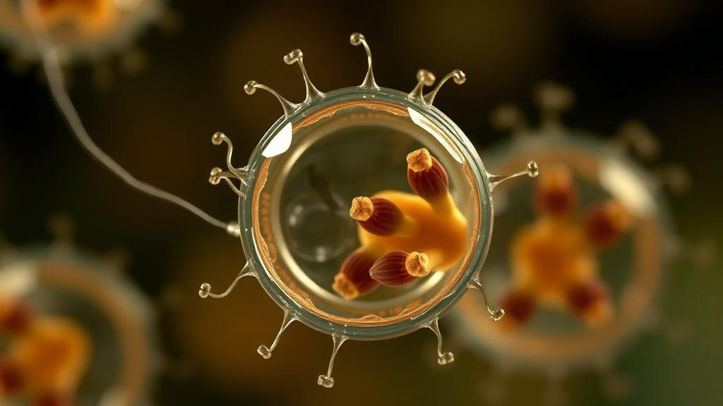
Cryptosporidium is one of the most consequential parasites in global health, a leading cause of diarrhoeal disease for which no fully effective drugs or vaccines exist. In otherwise healthy adults, infection causes self-limiting diarrhoea, but in young malnourished children and immunocompromised patients the disease can be severe, chronic and often fatal, contributing to wasting and growth stunting. The parasite infects a broad range of vertebrate hosts, and its complete life cycle unfolds within a single host, making both treatment and transmission control exceptionally difficult. Now, writing in PLOS Pathogens, researchers from the University of Dundee and Clemson University have synthesised what is known about one of the most neglected corners of this parasite’s biology: the female sexual stages, the cells that build the infectious oocyst and thereby drive every new infection.
The life cycle begins when a host ingests an oocyst, a resilient, wall-encased structure containing four sporozoites. In the gastrointestinal tract the oocyst excysts, releasing sporozoites that invade the epithelial cells lining the gut. Cryptosporidia are obligate intracellular parasites: they attach to the surface of a gut cell and pull the host membrane around themselves to form a parasitophorous vacuole, inside which they replicate. The sporozoite develops into a meront, duplicating its nucleus and organelles, and after roughly twelve hours divides into eight motile merozoites. These egress and invade neighbouring epithelial cells, repeating the asexual cycle. Remarkably, live-cell imaging of Cryptosporidium parvum infecting HCT-8 cells, together with single-cell RNA sequencing data from in vitro and in vivo infections, indicates that merozoites from the third round of asexual replication are committed directly to sexual development, apparently without a morphologically distinct type II meront of the kind described in other species such as C. muris and C. mortiferum.
Why and how the parasite commits to sexual stages after exactly three asexual rounds remains unknown. What is clear is that the committed merozoite invades a new epithelial cell and differentiates into either a macrogamont, which develops a single female gamete, or a microgamont, which produces sixteen male gametes. Mature males egress and seek out females, which never leave their host cell and instead mature intracellularly until fertilised. Fertilisation produces a zygote that undergoes meiosis, constructs the oocyst wall and completes sporogony to yield four haploid sporozoites within the protective shell. Because standard co-culture of C. parvum with HCT-8 cells does not support observation of fertilisation or subsequent zygote and oocyst development, sexual commitment, fertilisation, meiosis, sporogony and oocyst construction remain among the most mysterious phases of the life cycle.
Sexual commitment itself is mandatory for the third generation of merozoites and appears to be governed by an intrinsic, non-chromosomal mechanism. Live imaging has shown that merozoites from a single meront give rise to both sexes: of the eight merozoites in a meront, approximately two to three become male and the remaining five to six become female. This produces an excess of small, motile microgametes, presumably improving the odds that a female will be found and fertilised. One known player is the transcription factor Myb-M, expressed early in male development; experimental overexpression of Myb-M in C. parvum is sufficient to drive differentiation into males. No equivalent trigger for female commitment has been identified, leading the authors to suggest that female fate may be the default programme of sexual differentiation.
Female parasites, once committed, undertake one of the most demanding developmental programmes in the parasite’s repertoire. They carry a large nucleus, reflecting their status as the most transcriptionally and translationally active life cycle stage. Early in development, females express putative transporters, membrane proteins and secreted proteins hypothesised to mediate fertilisation, possibly facilitating male attachment and entry. Later, they mount two distinct waves of gene expression devoted to building the oocyst wall. The wall itself is a multilayered structure: an external glycocalyx, an outer layer of acid-fast lipids, and an inner layer of glycoproteins. These components are synthesised and stockpiled in female-exclusive organelles called Wall Forming Bodies, or WFBs, of which there are two subtypes distinguished by electron density. The prevailing hypothesis is that WFB Type I contents are secreted first to build the outer layer and WFB Type II contents follow to form the inner layer.
Only a handful of WFB components are confirmed. COWP1 is currently the only validated marker for WFB Type I, while COWP6 and COWP8 show punctate localisation in developing females, suggesting they also reside in these organelles. Transcriptomic and oocyst proteomic datasets have generated a comprehensive candidate list of WFB proteins, but how the organelles are synthesised, what exactly they contain, and how their secretion is regulated remain open questions. The single-cell atlas of C. parvum reveals that the first expression wave contains the Cryptosporidium Oocyst Wall Protein family and accessory enzymes such as proteases and protein modifiers for assembling the proteinaceous shell, while the second wave expresses glycosyltransferases and other secreted proteins including mucins and C-type lectins. These components are secreted into the space between the parasite plasma membrane and the host cell membrane, allowing the wall to be built while the parasite is still inside the gut cell. Having wall components ready just in time presumably keeps pace with the rapid turnover of epithelial host cells.
Females also provision their offspring. Genetic crosses using isolates tagged with different fluorescent proteins have shown that maternal cytoplasmic proteins persist in the sporozoites of first-generation progeny, including proteins involved in assembling dense granule organelles, which remodel the host cell after invasion. Females additionally express the proteins of the crystalloid body, a structure whose function is unknown but is likely involved in sporogony. Post-transcriptional control may be equally important: the maternal RNA-binding protein F-RBP, expressed in early females, binds female RNA transcripts, and conditional knockouts in vivo produce sterile sporozoites. F-RBP may therefore be essential for regulating maternally inherited RNA required to generate infectious sporozoites in the next generation.
Transcriptional regulation of female development is only beginning to be mapped. Two female-specific Apetala-2-type DNA-binding proteins have been identified: ApiAP2-1 is required for female differentiation, and ApiAP2-F is required for oocyst shedding. Insulinase-like protease 1 also contributes to female development and oocyst shedding. Intriguingly, females show reduced expression of ribosomal proteins, indicative of translational repression, and the authors hypothesise that epigenetic mechanisms such as chromatin remodelling may operate, consistent with the expansion of the female nucleus. Whatever the precise combination of mechanisms, they are fundamental to completing the life cycle and represent attractive targets for intervention.
What happens after fertilisation is even less understood. Fertilisation is presumed to trigger oocyst wall formation, sporogony and meiosis, but no culture system permits direct observation, so the signals regulating post-fertilisation development remain unknown across the coccidia. In Cryptosporidium, meiotic divisions appear to precede and partially overlap with deposition of the oocyst wall. Genes related to meiosis and DNA repair are expressed by females, including the DNA meiotic recombinase DMC1, detected in female nuclei. Under experimental conditions, C. parvum chromosomes show Mendelian inheritance with high frequencies of meiotic recombination, and frequent sex between genetically distinct parasites could accelerate the evolution and spread of traits such as virulence or drug resistance, as suggested by clofazimine resistance observed between C. parvum subtypes. Unlike other apicomplexans, Cryptosporidium oocysts are shed fully sporulated and immediately infectious, ready either to be excreted into the environment or to excyst in situ and reinfect the current host, a process that underpins the chronic disease seen in vulnerable patients.
A long-standing model proposes two oocyst types: thin-walled oocysts that excyst within the host to drive autoinfection, and thick-walled oocysts that are excreted and transmitted. The evidence for thin-walled oocysts rests on a few electron micrographs, and single-cell data show no transcriptional signature suggesting that some females are programmed to produce thin-walled oocysts. The authors instead hypothesise that all oocysts are thick-walled and environmentally robust, but that a small number excyst in the gut before excretion; given the exponential asexual cycle, even rare reinfections can sustain heavy parasite burdens. Improved culture systems, including organoid co-culture, are now making post-fertilisation biology experimentally accessible for the first time. Because oocyst biogenesis is exclusive to females and conserved across coccidia, dissecting this female-driven process could expose factors essential for transmission and open new avenues for drugs or vaccines. As the authors argue, it is time to stop overlooking Cryptosporidium females, the true engines of the parasite’s success.
Subject of Research: The biology of female sexual stages and oocyst formation in Cryptosporidium parasites
Article Title: Mysterious girls: The biology of female Cryptosporidium parasites
Article References: Stevens, S., Goddard, P., Walzer, K. A., & Pawlowic, M. C. (2026). Mysterious girls: The biology of female Cryptosporidium parasites. PLOS Pathogens, 22(10), e1014678. https://doi.org/10.1371/journal.ppat.1014678
Image Credits: AI Generated
DOI: 10.1371/journal.ppat.1014678
Keywords: Cryptosporidium, parasitology, oocyst, sexual development, gametogenesis, apicomplexa, Cryptosporidium parvum, transmission, diarrhoeal disease, Wall Forming Bodies, meiosis, global health
Cite Scienmag News
Kristina Jarvis. (October 13, 2026). The Hidden Lives of Female Cryptosporidium Parasites. Scienmag. https://scienmag.com/the-hidden-lives-of-female-cryptosporidium-parasites/
Kristina Jarvis. "The Hidden Lives of Female Cryptosporidium Parasites." Scienmag, 13 October 2026, https://scienmag.com/the-hidden-lives-of-female-cryptosporidium-parasites/. Accessed 13 October 2026.
Kristina Jarvis. "The Hidden Lives of Female Cryptosporidium Parasites." Scienmag. October 13, 2026. https://scienmag.com/the-hidden-lives-of-female-cryptosporidium-parasites/


