


For more than a century, the way doctors and biologists have looked at DNA inside cells has barely changed. A stain, a microscope, and a trained eye have been the tools of the trade, whether the sample is a routine hospital biopsy or a slice of tissue in a research laboratory. Now a team spanning Barcelona, Hong Kong and Guangdong has reported a fundamentally different way of seeing the genome: fluorescent molecules that blink on and off by themselves, allowing microscopes to resolve the packaging of DNA inside living cells at resolutions once thought impossible, and in preserved clinical tissue at a scale approaching the width of the DNA double helix itself.
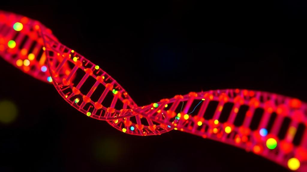
The work, published in the journal Molecular Cell, was led by researchers at the Centre for Genomic Regulation (CRG) in Barcelona together with the City University of Hong Kong and the Guangdong Provincial People’s Hospital of the Guangdong Academy of Medical Sciences at Southern Medical University. The team describes a new class of fluorescent probes, called HoTs, which are small enough to slip into living cells unaided and bind directly to DNA. What makes them special is their blinking behaviour: rather than glowing continuously, they flicker intermittently, like strings of fairy lights switching on and off at random.
That flickering is not a quirk but the key to the entire technique. Conventional light microscopes hit a hard physical limit at roughly 200 nanometres, the diffraction barrier that blurs anything packed more tightly than that. If every dye molecule in a sample lit up simultaneously, even the most advanced microscope would simply record an overlapping glow. But if molecules emit one at a time, or in small sparse groups, each flash can be localised individually. Thousands of snapshots, each capturing a scattered subset of blinking probes, can then be stitched together by software into a reconstructed image far sharper than any single exposure. This is the principle behind single-molecule localisation microscopy, and the new probes were engineered specifically to deliver the right blinking dynamics for it.
With the HoT dyes, the researchers deployed STORM, a stochastic optical reconstruction microscopy technique, to visualise DNA inside living human cells at a resolution of 20 nanometres. That is a tenfold improvement over the conventional limit, achieved not on fixed and chemically treated samples but on cells that were alive throughout the imaging. The team demonstrated the approach in living human skin cells and in living HeLa cancer cells grown in the laboratory. In parallel, they pushed the technology even further on preserved cells, using MINFLUX, an ultra-high-resolution method that tracks individual fluorophores, to pinpoint single dye molecules to within three nanometres. The diameter of the DNA double helix is approximately two nanometres, so a localisation precision of three nanometres brings imaging to the very doorstep of the molecule itself.
The significance of that achievement becomes clear when one considers what biologists have been missing. Every human cell contains roughly two metres of DNA, folded and compressed into a nucleus only a few micrometres across. The degree to which that DNA is wound tightly or left loose is not decorative; it is one of the principal layers of biological control. Tightly packed DNA tends to be silent, while loosely packed regions are accessible to the molecular machinery that reads genes. Yet almost all existing images of DNA folding come from dead, fixed cells, because the powerful microscopy techniques needed to track individual genomic components typically require harsh chemicals and intense laser illumination that living cells cannot survive. The new probes sidestep that constraint, allowing chromatin, the natural state of DNA in cells, to be watched in motion.
Professor Hongyan Sun of the City University of Hong Kong, co-senior author of the study, highlighted the central design problem: the key challenge was to create fluorophores with the right blinking behaviour for super-resolution imaging. ICREA Research Professor Pia Cosma of the CRG, senior author, explained the dual power of the system: with the same dye, researchers can watch DNA moving in a living cell, revealing how chromatin behaves, and zoom into preserved cells until they are almost at the scale of the DNA molecule itself. Combining both approaches, she noted, allows one of the main layers of control in human biology to be seen at unprecedented resolution.
The team did not stop at laboratory cell lines. In a striking demonstration of clinical relevance, they applied the probes to ordinary wax-preserved tissue slices taken from three bowel cancer patients, the same kind of samples hospitals routinely store after biopsies. In the tumour tissue, the DNA was noticeably looser and more spread out than in the healthy tissue sitting immediately beside it. This observation aligns with earlier findings that DNA unpacks progressively as cancer takes hold, and it suggests a provocative possibility: how loosely a cell’s DNA is folded could eventually serve as a measurable indicator of how far a tumour has progressed or how aggressive it is. Today, doctors examine these biopsies largely by eye using staining methods more than a century old. The ability to add a three-dimensional readout of how DNA occupies space inside cells could one day become an additional diagnostic clue.
The probes also proved useful beyond human tissue. The researchers imaged slices of zebrafish eye, an animal famous for its ability to regrow damaged retina. Retinal regeneration in these fish is thought to involve cells loosening their DNA packaging in order to become flexible again, and the new dyes offer a way to observe that process directly at nanometre scale. Such experiments hint at a broader future for the technology in regenerative medicine, where identifying which cells have entered a plastic, stem-like state is a central challenge.
Artificial intelligence enters the story as a force multiplier. The same team had previously unveiled a program called AINU, short for AI of the Nucleus, in 2024, which scans super-resolution images of cellular DNA and detects patterns far too subtle for the human eye. Originally, AINU could only analyse cells that had been killed and preserved. Trained on the new living-cell images produced with the HoT probes, the AI correctly distinguished skin cells from stem cells between 96 and 98 per cent of the time. The two cell types carry identical genomes but fold their DNA differently, and the algorithm could see that difference in living material. The researchers hope the same principle could eventually be turned to distinguishing cancerous from healthy tissue, or to selecting the most promising stem cells to accelerate regenerative medicine.
The technology is not without limits, and the authors are candid about them. The probes coat the entirety of the DNA rather than highlighting any single gene, so they reveal global packaging architecture rather than gene-specific organisation. The very sharpest pictures, at the three-nanometre scale, required preserved rather than living cells, although the team reports that follow-up studies are already under way and that MINFLUX has the potential to image the probes in living cells as well. Even so, the combination demonstrated here, live-cell chromatin dynamics at 20 nanometres and near-helix-scale localisation in preserved clinical samples, marks a genuine step change. A staining method from the nineteenth century may soon share the microscope bench with blinking molecular probes and AI readers, giving clinicians and researchers a view of the genome not as a static blueprint but as a living, breathing three-dimensional structure whose shape itself carries diagnostic information.
Subject of Research: Super-resolution fluorescence imaging of DNA packaging in living and preserved cells using self-blinking probes
Article Title: Self-blinking ‘fairy lights’ allow DNA to be imaged at almost double helix width resolution
Article References: Self-blinking ‘fairy lights’ allow DNA to be imaged at almost double helix width resolution. (n.d.). Original publication
Image Credits: AI Generated
DOI: Not provided
Keywords: super-resolution microscopy, STORM, MINFLUX, chromatin, DNA packaging, fluorescent probes, cancer diagnostics, biopsy imaging, artificial intelligence, stem cells, zebrafish, Molecular Cell
Cite Scienmag News
Grant Pearson. (October 11, 2026). Blinking fluorescent probes reveal DNA packaging in living cells at near-helix resolution. Scienmag. https://scienmag.com/blinking-fluorescent-probes-reveal-dna-packaging-in-living-cells-at-near-helix-resolution/
Grant Pearson. "Blinking fluorescent probes reveal DNA packaging in living cells at near-helix resolution." Scienmag, 11 October 2026, https://scienmag.com/blinking-fluorescent-probes-reveal-dna-packaging-in-living-cells-at-near-helix-resolution/. Accessed 11 October 2026.
Grant Pearson. "Blinking fluorescent probes reveal DNA packaging in living cells at near-helix resolution." Scienmag. October 11, 2026. https://scienmag.com/blinking-fluorescent-probes-reveal-dna-packaging-in-living-cells-at-near-helix-resolution/


