


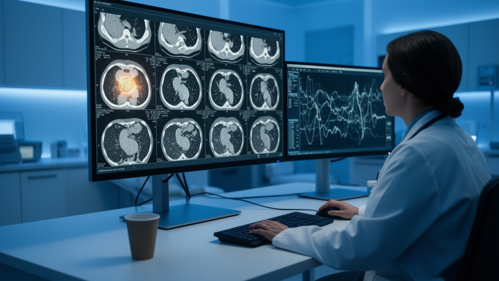
A routine CT scan already sits in the hospital record of nearly every patient diagnosed with a gastrointestinal stromal tumor, the most common mesenchymal tumor of the digestive tract. What if that same scan, without a single additional needle stick, could reveal how aggressively the tumor is likely to behave? A new systematic review and meta-analysis published in BMC Medical Imaging suggests that artificial intelligence algorithms trained to extract invisible patterns from contrast-enhanced CT images can indeed predict the status of Ki-67, a pivotal marker of cell proliferation, with moderate accuracy. The findings, compiled by a large multidisciplinary team of radiologists, pathologists, and evidence-based medicine researchers, offer both a promising step toward non-invasive tumor profiling and a sobering reminder of how far the field still must travel before such tools reach the clinic.
Ki-67 is a protein that appears in cells whenever they are actively dividing. Pathologists stain biopsy tissue for it and report the percentage of positive nuclei, a number known as the proliferation index. In gastrointestinal stromal tumors, or GISTs, this index is woven directly into risk stratification schemes that guide whether a patient needs close surveillance or preventive drug therapy with tyrosine kinase inhibitors. The problem is that obtaining the index requires tissue, usually acquired through endoscopic biopsy or full surgical resection. Biopsies of GISTs can be technically difficult, may not sample the most proliferative regions of a heterogeneous tumor, and in some cases carry a small risk of tumor rupture or seeding. A reliable way to estimate Ki-67 from imaging alone would therefore fill a genuine clinical gap, allowing risk assessment to begin at the moment the tumor is first seen on a scan.
The technique at the heart of the new analysis is radiomics, a branch of medical imaging informatics that converts the pixels of a medical image into hundreds of quantitative features. Where a radiologist’s eye perceives a mass with irregular borders and heterogeneous enhancement, a radiomics pipeline measures first-order statistics describing the distribution of gray values, texture features capturing spatial relationships between neighboring voxels, and higher-order descriptors derived from mathematical transforms such as wavelets. These features, often combined with clinical variables and processed by machine learning classifiers, can encode subtle biological properties of a tumor, including its cellularity, necrosis, and mitotic activity, that correlate with proliferation. Because contrast-enhanced CT is already the standard staging examination for GISTs, radiomics models built on these scans require no new imaging, no new cost, and no new risk to the patient.
To determine how well such models actually perform, the research team followed the PRISMA-DTA reporting guideline and systematically searched five major databases, including PubMed, Embase, Scopus, Web of Science, and the Chinese CNKI database, for studies published through October 20, 2025. Their search identified eleven retrospective studies encompassing 2,868 patients and eleven validation cohorts. Only four of the studies reported external validation, meaning that most models were tested on data drawn from the same institutions where they were developed. The quality of each study was appraised with QUADAS-2, the standard tool for diagnostic accuracy research, while the methodological rigor of the radiomics pipelines themselves was scored with the METRICS instrument, a checklist designed specifically to expose weaknesses in machine learning imaging studies.
The statistical centerpiece of the analysis was a bivariate random-effects model, the preferred approach for pooling paired sensitivity and specificity estimates across studies. The headline numbers were striking in their asymmetry. Across all validation cohorts, contrast-enhanced CT radiomics models achieved a pooled sensitivity of 0.67 for distinguishing tumors with high Ki-67 expression from those with low expression, but a pooled specificity of 0.82. The summary area under the receiver operating characteristic curve, a single number summarizing overall discriminative ability, came to 0.75. In practical terms, the models were considerably better at correctly identifying slow-proliferating tumors than at catching the aggressive ones. A specificity of 0.82 means that roughly four out of five tumors flagged as low risk truly were low risk, but a sensitivity of 0.67 means that about a third of genuinely high-proliferation tumors would slip through undetected if the model were used alone.
That asymmetry matters enormously for the intended clinical use. If a radiomics score were deployed as a triage tool to reassure clinicians that a small GIST could simply be watched, the tumors it misses would be precisely the ones most likely to metastasize. Conversely, the relatively strong specificity suggests the models could be valuable as a rule-in test, flagging tumors that warrant expedited surgery or neoadjuvant therapy. The authors of the meta-analysis frame the technology accordingly: as a potential adjunct to, not a replacement for, conventional risk stratification. Read alongside tumor size, location, and mitotic count, an imaging-derived proliferation estimate could sharpen preoperative decision-making even at its current level of accuracy.
Deeks’ funnel plot asymmetry test, a statistical check for small-study effects in diagnostic meta-analyses, returned a p-value of 0.63, providing no evidence of significant publication bias in the pooled estimates. This is reassuring, because diagnostic accuracy literature is notoriously prone to the file drawer problem, in which studies with disappointing results go unpublished. The heterogeneity picture was less comforting. Specificity estimates varied substantially across studies, a statistical scatter that the bivariate model must absorb and that signals genuine differences in how the underlying models were built. Sources of that variability are easy to imagine: different CT scanners and reconstruction kernels, different segmentation strategies ranging from manual contouring to semi-automated approaches, different feature selection methods, and different classifiers ranging from logistic regression to support vector machines and random forests.
Two prespecified subgroup analyses probed the moderators most likely to drive that heterogeneity. The first compared studies using different Ki-67 cut-offs to define high proliferation, with some studies drawing the line at 5 to 6 percent and others at 8 to 10 percent. Perhaps surprisingly, the differences between these thresholds were small, suggesting that the radiomics signal for proliferation is robust enough to survive reasonable variation in how the ground truth is defined. The second subgroup examined the software used to extract features. Studies built on PyRadiomics, the widely used open-source radiomics platform, showed lower specificity than those using other tools. The authors are careful to stress that these comparisons rest on very few studies per subgroup and should be interpreted cautiously, but the finding is a useful flag for the field: standardization of the computational pipeline itself, not just the clinical endpoint, may shape real-world performance.
The limitations that temper enthusiasm are laid out with unusual candor. Every included study was retrospective, meaning the models were trained and tested on patients whose outcomes were already known, an arrangement that almost always flatters performance relative to prospective use. Only a minority of studies achieved external validation on independent cohorts, and none of the evidence base spans multiple countries or centers in a coordinated way. Radiomics features are notoriously sensitive to scanner vendor, acquisition protocol, and image preprocessing, so a model calibrated on one institution’s CT fleet may degrade when exported elsewhere. The QUADAS-2 and METRICS assessments, while not detailed in the summary results, exist precisely because methodological shortcuts, such as inadequate sample sizes per feature, optimistic feature selection performed on the full dataset, and absent reporting of model tuning, are endemic in radiomics research and can inflate apparent accuracy.
What would it take for CT-based Ki-67 prediction to earn a place in clinical guidelines? The authors point squarely to prospective, multinational validation studies: models locked before use, tested on consecutive patients at centers that did not participate in development, with standardized imaging protocols and harmonized Ki-67 thresholds. Until then, the meta-analysis delivers a measured verdict that is itself a kind of milestone. Radiomics has moved from proof-of-concept papers to a pooled evidence base spanning nearly three thousand patients, with a summary AUC of 0.75 that is respectable for a purely imaging-derived biomarker of a molecular phenotype. For patients with GISTs, the vision is compelling: the scan they already receive, read by an algorithm that has learned to see proliferation, flagging aggressive biology before the first incision is made. The new analysis shows that vision is technically plausible and statistically grounded, while insisting, correctly, that the last mile of clinical translation has not yet been walked.
Subject of Research: Diagnostic performance of contrast-enhanced CT-based radiomics models for predicting Ki-67 proliferation status in gastrointestinal stromal tumors
Article Title: Diagnostic performance of contrast-enhanced CT-based radiomics models for Ki-67 status prediction in gastrointestinal stromal tumors: a systematic review and meta-analysis
Article References: Diagnostic performance of contrast-enhanced CT-based radiomics models for Ki-67 status prediction in gastrointestinal stromal tumors: a systematic review and meta-analysis. (n.d.). https://doi.org/10.1186/s12880-026-02769-3
Image Credits: AI Generated
DOI: 10.1186/s12880-026-02769-3
Keywords: gastrointestinal stromal tumors, Ki-67, radiomics, contrast-enhanced CT, meta-analysis, diagnostic accuracy, machine learning, medical imaging, cancer imaging, risk stratification, predictive medicine, BMC Medical Imaging
Cite Scienmag News
Ophelia Keating. (October 8, 2026). AI Reads Routine CT Scans to Predict Aggressiveness of Stomach and Gut Tumors. Scienmag. https://scienmag.com/ai-reads-routine-ct-scans-to-predict-aggressiveness-of-stomach-and-gut-tumors/
Ophelia Keating. "AI Reads Routine CT Scans to Predict Aggressiveness of Stomach and Gut Tumors." Scienmag, 8 October 2026, https://scienmag.com/ai-reads-routine-ct-scans-to-predict-aggressiveness-of-stomach-and-gut-tumors/. Accessed 8 October 2026.
Ophelia Keating. "AI Reads Routine CT Scans to Predict Aggressiveness of Stomach and Gut Tumors." Scienmag. October 8, 2026. https://scienmag.com/ai-reads-routine-ct-scans-to-predict-aggressiveness-of-stomach-and-gut-tumors/


