


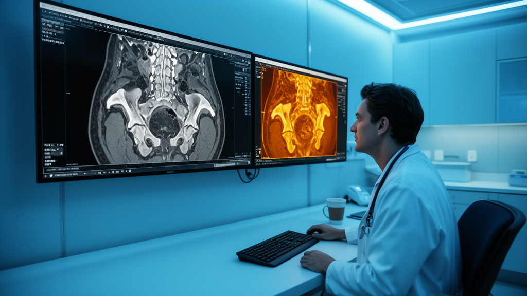
Prostate cancer screening has long been caught in an uncomfortable trade-off. Men with elevated prostate-specific antigen levels or suspicious digital rectal examinations often undergo needle biopsies that turn out to be negative, while some tumors that truly need treatment can be missed or underestimated. A new study published in BMC Medical Imaging by researchers at the Second People’s Hospital of Nantong in Jiangsu Province, China, offers a way to sharpen that decision. The team, led by Yang Lv and corresponding author Haiyan Yuan, built a machine-learning model that fuses standard clinical information, radiologist scores, and quantitative image features extracted from biparametric MRI to predict, before any needle is inserted, whether a patient harbors clinically significant prostate cancer.
The distinction at the heart of the study is between prostate cancer in general and clinically significant prostate cancer, often abbreviated csPCa. Many prostate tumors are indolent: they grow so slowly that they would never threaten a man’s life, and guidelines increasingly favor active surveillance over immediate treatment for such cases. Clinically significant disease, by contrast, is aggressive enough to warrant biopsy confirmation and possible intervention. Separating the two non-invasively is one of the most consequential problems in urologic oncology, because it determines who needs an invasive procedure and who can safely be monitored. The Nantong team’s goal was to build a tool that makes that separation more reliable than current practice allows.
Their approach rests on biparametric MRI, a protocol that uses two sequences rather than the full multiparametric set. The first is T2-weighted imaging, which reveals the anatomy of the prostate gland and any structural abnormalities within it. The second is the apparent diffusion coefficient map, derived from diffusion-weighted imaging, which measures how freely water molecules move through tissue. Cancerous tissue is typically denser and more cellular than healthy tissue, restricting water movement and producing characteristic dark regions on ADC maps. Because biparametric MRI skips the dynamic contrast-enhanced sequence, it requires no gadolinium injection, shortens scan time, and lowers cost, which has made it increasingly attractive for widespread prostate cancer detection programs.
On top of these standard images, the researchers layered radiomics, a technique that converts medical images into high-dimensional quantitative data. Using the open-source 3D Slicer platform, the team delineated regions of interest on both the T2-weighted images and the ADC maps for each patient. They then used PyRadiomics, a widely adopted open-source library, to extract hundreds of mathematical features from each region: statistics describing the distribution of pixel intensities, measures of texture such as entropy and homogeneity, and shape descriptors capturing the size and irregularity of suspicious areas. The premise is that these features encode subtle tissue properties invisible to the human eye, providing a quantitative fingerprint of the tumor microenvironment.
Raw radiomics data, however, come with a statistical hazard: far more features than the 458 patients in the study, which invites overfitting, the phenomenon in which a model memorizes the quirks of its training data rather than learning generalizable patterns. To control this, the researchers applied dimensionality reduction followed by least absolute shrinkage and selection operator regression, known as LASSO. LASSO works by penalizing the model for using too many variables, effectively shrinking the coefficients of uninformative features to zero and retaining only the most predictive subset. This step is essential for radiomics studies, because without it, models routinely achieve dazzling internal performance that collapses when confronted with new patients.
The study population of 458 men, all with complete clinical and biparametric MRI data, was randomly divided in a 7:3 ratio into a training set and a validation set. The researchers built three tiers of models. The first was a clinical model using conventional variables such as age, prostate-specific antigen level, prostate volume, and the PI-RADS score, the standardized five-point scale radiologists assign to MRI findings based on the likelihood of clinically significant cancer. The second was a radiomics-only model. The third combined everything: clinical variables, PI-RADS scores, and the selected radiomics features. For each tier, they compared four machine-learning algorithms: logistic regression, support vector machine, random forest, and XGBoost, a gradient-boosting method popular in machine-learning competitions.
The results, reported in the validation cohort, tell a clear story. The clinical model alone achieved an area under the receiver operating characteristic curve, or AUC, of 0.843, meaning it distinguished significant from insignificant disease reasonably well. The radiomics logistic regression model reached 0.888. The combined logistic regression model topped both at 0.900, and the improvement was statistically significant: p less than 0.001 against the clinical model and p equal to 0.032 against the radiomics model. The combined model also achieved a sensitivity of 0.841, correctly flagging the large majority of men with clinically significant cancer, and showed strong calibration, with a calibration slope of 0.950, indicating that its predicted probabilities closely matched observed outcomes.
Notably, the team did not simply crown the model with the highest AUC. They evaluated all candidates across three dimensions: discrimination, calibration, and clinical utility, the last assessed with decision curve analysis across a threshold probability range of 10 to 60 percent. Decision curve analysis asks a practical question: at what level of suspicion would a clinician act, and does the model produce more benefit than harm across that range? After weighing all three criteria, the combined logistic regression model emerged as the winner, and the researchers translated it into a nomogram, a graphical scoring tool that lets a clinician plot individual patient values on simple axes and read off a total risk score. The nomogram demonstrated good calibration and consistent net benefit across the clinically relevant threshold range.
The practical implications are substantial. A urologist facing a patient with a borderline PI-RADS 3 lesion and mildly elevated PSA could enter the patient’s data into the nomogram and obtain a quantified probability of clinically significant cancer. For men whose scores fall well below the biopsy threshold, the tool supports a strategy of surveillance and repeat imaging rather than immediate needle sampling, potentially sparing them the pain, bleeding, infection risk, and anxiety of an unnecessary procedure. For men with high scores, it reinforces the case for prompt biopsy. Because the model relies only on biparametric MRI and routine clinical data, it requires no contrast agent, no additional scanning, and no new equipment beyond the software pipeline, making it realistic to deploy in hospitals that already perform prostate MRI.
The authors and outside observers alike caution that this is a retrospective, single-center study, and the model will need external validation on independent cohorts at other institutions before it can change clinical guidelines. The ethics committee of Nantong Second People’s Hospital approved the work, which was funded by the Special Research Project of the Nantong Municipal Health Commission, and the article is published open access under a Creative Commons license. Still, the study adds to a fast-growing body of evidence that radiomics and machine learning can extract diagnostic value from images that radiologists already acquire every day. If validated more broadly, this kind of nomogram could become a standard checkpoint between the MRI scanner and the biopsy gun, ensuring that needles are directed where they matter and held back where they do not.
Subject of Research: A radiomics-clinical nomogram based on biparametric MRI for risk stratification of clinically significant prostate cancer
Article Title: A radiomics-clinical nomogram based on biparametric MRI for improved risk stratification of clinically significant prostate cancer
Article References: A radiomics-clinical nomogram based on biparametric MRI for improved risk stratification of clinically significant prostate cancer. (n.d.). https://doi.org/10.1186/s12880-026-02888-x
Image Credits: AI Generated
DOI: 10.1186/s12880-026-02888-x
Keywords: prostate cancer, biparametric MRI, radiomics, machine learning, nomogram, PI-RADS, clinical decision support, medical imaging, LASSO regression, biopsy, predictive medicine, cancer imaging
Cite Scienmag News
Nathaniel Bowman. (October 8, 2026). AI Reads MRI Scans to Spot Dangerous Prostate Cancer Before Biopsy. Scienmag. https://scienmag.com/ai-reads-mri-scans-to-spot-dangerous-prostate-cancer-before-biopsy/
Nathaniel Bowman. "AI Reads MRI Scans to Spot Dangerous Prostate Cancer Before Biopsy." Scienmag, 8 October 2026, https://scienmag.com/ai-reads-mri-scans-to-spot-dangerous-prostate-cancer-before-biopsy/. Accessed 8 October 2026.
Nathaniel Bowman. "AI Reads MRI Scans to Spot Dangerous Prostate Cancer Before Biopsy." Scienmag. October 8, 2026. https://scienmag.com/ai-reads-mri-scans-to-spot-dangerous-prostate-cancer-before-biopsy/


