


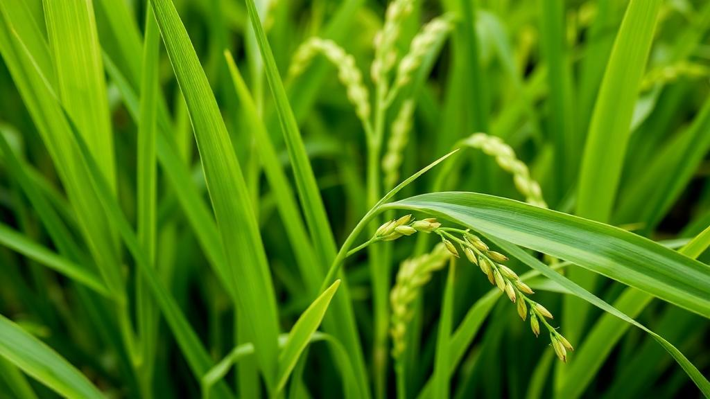
A single protein made by rice grassy stunt virus can restructure the physical architecture of the rice plant, according to a study published in the journal Stress Biology. The protein, known as p2, is encoded by the second RNA segment of the virus, a tenuivirus that spreads through rice fields in Southeast Asia and is transmitted by the brown planthopper, Nilaparvata lugens. When researchers engineered rice plants to overproduce p2, the plants grew shorter, developed brittle leaves and stems, and became markedly more vulnerable to several unrelated viruses. Yet the same plants turned out to be significantly more resistant to the brown planthopper, the very insect that carries the virus from plant to plant. The finding reveals an unexpected trade-off in which a viral protein simultaneously opens the door to viral infection and closes it to the vector that delivers the pathogen.
The plant cell wall is far more than a passive scaffold. Composed chiefly of cellulose, hemicellulose and pectin, it provides mechanical support, determines plant height and stem strength, and acts as a first line of defense against invading pathogens. Previous genome-wide studies had shown that infection by rice grassy stunt virus, as well as by rice dwarf virus and rice tungro spherical virus, suppresses the expression of genes involved in cell wall biosynthesis, including genes encoding cellulose synthases and arabinogalactan proteins. What remained unclear was which viral component was responsible and how it interfered with wall construction. The new work, led by researchers at Fujian Agriculture and Forestry University, points to p2 as a key player in this process.
To dissect the protein’s function, the team cloned the full-length RNA2 sequence of the virus and introduced it into the rice cultivar Nipponbare, generating three independent transgenic lines that overexpress p2. Compared with wild-type plants, all three lines were shorter, produced fewer productive panicles, and had lower thousand-grain weights. Microscopic examination of leaf sheath cells showed that cell length was significantly reduced, indicating that p2 expression suppresses cell expansion during development. The uppermost and second-uppermost internodes were notably shorter than in controls, consistent with the stunting that characterizes grassy stunt disease in the field.
The most striking phenotype, however, was brittleness. Mechanical testing with a micro-force testing device showed that the elongation and breaking forces of leaves from the p2-overexpressing plants were reduced by 78 and 85 percent respectively, relative to wild type. For the second internodes, these forces fell by 80 and 77 percent. Crucially, plants naturally infected with rice grassy stunt virus showed the same tendency: their leaves lost 54 percent of elongation force and 62 percent of breaking force, while internodes lost 23 and 26 percent. The overlap between the transgenic and the infected phenotypes strongly suggests that p2 is a principal driver of the brittle symptom seen during natural infection.
Scanning electron microscopy explained the mechanical weakness. The sclerenchyma cell walls, the thickened support tissues of leaves and stems, were significantly thinner in the p2-expressing plants than in wild type. Biochemical assays quantified the compositional shift: cellulose content dropped significantly while lignin accumulated to markedly higher levels in both leaves and stems. Calcofluor white staining, which labels cellulose, confirmed the reduction in that polymer under the fluorescence microscope. When the researchers examined cross-sections of virus-infected rice, they saw a more relaxed cell wall architecture and the same pattern of reduced cellulose and elevated lignin, mirroring the transgenic lines and reinforcing the link between p2 and wall remodeling during infection.
Where does the protein act? Transient expression assays in Nicotiana benthamiana showed that p2 localizes mainly to the cytoplasm and the nucleus. But RNA in situ hybridization on infected rice tissues revealed strong p2 transcript signals in fundamental and mechanical tissues, hinting at an association with structural structures. The team then separated infected leaf homogenates into four subcellular fractions: cell wall, organelle-enriched, crude membrane, and soluble cytoplasmic. Immunoblotting showed that while most p2 sat in the soluble fraction, a detectable amount appeared in the cell wall fraction, which was validated by the enrichment of the known wall-associated protein BC1. A control viral protein, P3, was found only in the soluble fraction and never in the wall. Plasmolysis experiments in tobacco epidermal cells added further support: after treatment with concentrated salt solution, free GFP retracted fully into the protoplast, whereas a p2-GFP fusion retained partial co-localization with the cell wall marker EXPA8-mCherry at the cell periphery. Together, these data indicate a small but reproducible pool of p2 that physically associates with the wall.
Transcriptome profiling of 42-day-old plants clarified the molecular consequences. RNA sequencing identified 1,811 upregulated and 1,574 downregulated genes in the p2-overexpressing lines. Downregulated genes were significantly enriched in protein processing in the endoplasmic reticulum, plant-pathogen interactions, and amino sugar and nucleotide sugar metabolism, all pathways tied to cell wall biosynthesis and immunity. Among the suppressed genes were members of an NAD-dependent epimerase/dehydratase family, GHMP kinase ATP-binding proteins, and the protein disulfide isomerase OsPDIL1-4, each previously implicated in sugar metabolism, protein folding, or acetylation of structural macromolecules essential for wall assembly. Upregulated genes clustered in ribosome, plant hormone signal transduction, and cofactor biosynthesis pathways, suggesting compensatory responses to disrupted growth. Notably, OsEBF2, a negative regulator of ethylene signaling known to promote resistance to the brown planthopper, was among the upregulated genes, providing a possible molecular thread connecting p2 expression to insect resistance.
The ecological consequences of this remodeling proved to be double-edged. In host preference assays, brown planthoppers settled on p2-overexpressing plants in significantly lower numbers beginning at 48 hours after release. Oviposition was markedly reduced, honeydew excretion, a proxy for feeding activity, was significantly lower, and survival assays showed that the transgenic plants survived insect inoculation better than both wild type and the susceptible variety TN1 at eight days after infestation. The altered wall stiffness, lignin deposition, or sugar metabolism appears to change the physical and nutritional landscape of the tissues, impairing the insect’s access to phloem sap or its feeding efficiency. In sharp contrast, the same plants were more susceptible to viruses. When inoculated with RGSV-carrying planthoppers, all three transgenic lines showed symptoms by four weeks and accumulated higher levels of viral RNAs and proteins than infected wild-type plants, with higher infection rates across multiple time points. The effect was not limited to the virus that produces p2: the plants also developed more severe symptoms and higher viral loads after inoculation with rice dwarf virus and Southern rice black-streaked dwarf virus, two double-stranded RNA viruses, indicating broad-spectrum susceptibility.
To test whether cell wall defects alone could explain this vulnerability, the researchers turned to two classic brittle culm mutants of rice. The bc1 mutant carries a mutation in a COBRA-like gene expressed in sclerenchyma cells and vascular bundles, resulting in thinner walls, reduced cellulose, and increased lignin. The bc13 mutant harbors a missense mutation in the cellulose synthase gene OsCESA9 that reduces mechanical strength to roughly one-third of wild type and cuts cellulose content by 22 percent. When inoculated with rice grassy stunt virus, both mutants developed more pronounced stunting than infected wild-type plants, accumulated significantly higher levels of viral RNAs and proteins, and showed higher infection rates over time. Both mutants also displayed altered brown planthopper responses compared with wild type. These results independently confirm that weakened cell walls facilitate viral invasion while modifying insect interactions, supporting the hypothesis that wall integrity is a genuine barrier to virus infection rather than an incidental correlate.
The study positions p2 as a novel structural effector, a viral protein that does not merely suppress immune signaling but physically undermines the host’s mechanical defenses. It also raises intriguing evolutionary questions. Rice grassy stunt virus depends on the brown planthopper for transmission, yet the wall weakening that benefits viral infection appears to hinder the vector’s colonization and feeding. Such a feedback loop could constrain transmission opportunities and shape virus evolution in ways not dictated solely by host compatibility. Beyond pathology, the findings carry agronomic and industrial implications. Brittle culm mutants are known to have improved straw palatability and digestibility for livestock and enhanced suitability for bioethanol production, so careful manipulation of cell wall traits could serve crop protection, feed quality, and renewable energy goals simultaneously. The authors propose that cell wall integrity should be viewed as a dual-purpose trait, governing both disease susceptibility and ecological fitness, and their work offers a mechanistic entry point for breeding rice varieties that balance these competing demands.
Subject of Research: How the rice grassy stunt virus p2 protein remodels rice cell wall integrity to alter virus susceptibility and brown planthopper resistance
Article Title: A viral protein disrupts rice cell wall integrity and modulates interactions with viruses and insects
Article References: Zhang, J., Ye, W., Li, S., Dong, C., Peng, F., Wang, C., Qin, J., Hu, Q., Zhang, J., Wu, J., & Zhao, S. (2026). A viral protein disrupts rice cell wall integrity and modulates interactions with viruses and insects. Stress Biology, 6(1), Article 17. https://doi.org/10.1007/s44154-026-00287-4
Image Credits: AI Generated
DOI: 10.1007/s44154-026-00287-4
Keywords: rice grassy stunt virus, p2 protein, cell wall, cellulose, lignin, brittle culm, brown planthopper, tenuivirus, plant virology, transcriptomics, plant immunity, Oryza sativa
Cite Scienmag News
Kristina Jarvis. (October 4, 2026). Viral Protein Weakens Rice Cell Walls, Boosting Virus Infection but Blocking Insect Pests. Scienmag. https://scienmag.com/viral-protein-weakens-rice-cell-walls-boosting-virus-infection-but-blocking-insect-pests/
Kristina Jarvis. "Viral Protein Weakens Rice Cell Walls, Boosting Virus Infection but Blocking Insect Pests." Scienmag, 4 October 2026, https://scienmag.com/viral-protein-weakens-rice-cell-walls-boosting-virus-infection-but-blocking-insect-pests/. Accessed 4 October 2026.
Kristina Jarvis. "Viral Protein Weakens Rice Cell Walls, Boosting Virus Infection but Blocking Insect Pests." Scienmag. October 4, 2026. https://scienmag.com/viral-protein-weakens-rice-cell-walls-boosting-virus-infection-but-blocking-insect-pests/


