


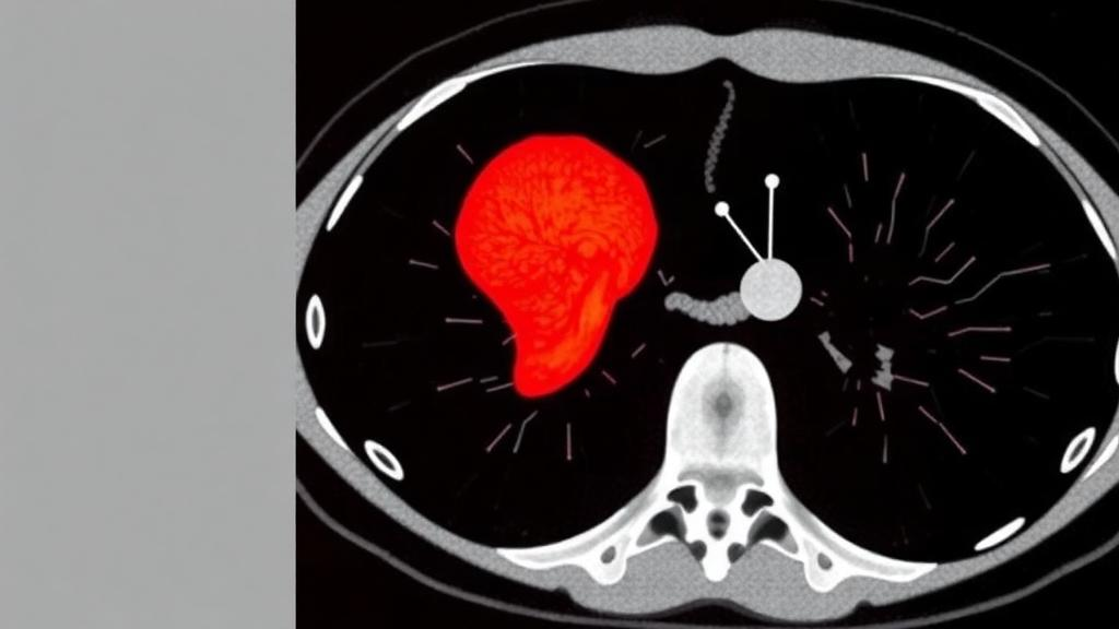
A small lump on the adrenal gland is one of the most common accidental findings in modern medicine. Radiologists call them incidentalomas, and they turn up in a striking fraction of abdominal CT scans performed for entirely unrelated reasons. Most of these nodules are benign adenomas, harmless growths that will never bother the patient. But a meaningful minority are metastases, fragments of cancer that have spread from elsewhere in the body, most often from the lung, breast, kidney or the melanoma of the skin. The difference between the two diagnoses can determine whether a patient undergoes surveillance, surgery or systemic cancer treatment, and telling them apart non-invasively has long been one of the stubborn problems of abdominal imaging.
The classic trick that radiologists use relies on fat. Typical adrenal adenomas are packed with intracellular lipid, which makes them lose signal on dedicated CT protocols and allows a confident benign diagnosis. But a substantial share of adenomas, known as lipid-poor adenomas, do not contain enough fat to betray themselves this way. On a conventional scan they look disconcertingly similar to metastases, and neither size, shape nor enhancement pattern can reliably separate them. The result is a diagnostic gray zone in which many patients end up needing biopsy, additional imaging with PET or chemical-shift MRI, or surgical removal of a gland that may have been perfectly healthy.
A new study published in BMC Medical Imaging suggests that the answer may already be hiding inside the images clinicians routinely acquire. A team led by Fangmei Zhu and Jian Wang, working across hospitals affiliated with Bengbu Medical University and Zhejiang Chinese Medical University, built machine learning models that read the invisible texture of adrenal tumors on multiphasic contrast-enhanced CT scans. The approach, known as radiomics, converts the pixel-level patterns of a medical image into hundreds of quantitative features describing texture, intensity distribution, coarseness and spatial relationships, features far too subtle for the human eye to grade but readily digestible by algorithms.
The researchers assembled a retrospective cohort of 425 patients, 212 with pathologically confirmed adrenal metastases and 213 with lipid-poor adenomas, drawn from two hospitals. Images from the First Affiliated Hospital of Bengbu Medical University, comprising 178 metastases and 189 adenomas, were used to train and internally validate the models, while a completely separate set of 58 patients from Tongde Hospital of Zhejiang Province served as an external validation cohort, the gold standard for testing whether a model has learned genuine biology rather than the quirks of a single scanner or population. Radiomic features were extracted separately from three phases of the CT examination: the unenhanced scan, the arterial phase captured shortly after contrast injection, and the venous phase acquired a little later.
From each phase the team derived features drawn from established texture families, including gray level co-occurrence matrices, run length and size zone matrices, and neighborhood gray-tone difference measures, each capturing a different aspect of how pixel intensities are arranged within the tumor. After dimensionality reduction and feature selection using techniques such as LASSO and multi-cluster feature selection, the researchers trained four different classifiers: Adaptive Boosting, Decision Tree, K-Nearest Neighbors and Logistic Regression. They then compared these against a conventional baseline model built from routine clinical and radiological indicators, the kind of information a radiologist weighs in everyday practice, and also tested a fusion model that combined features from all three CT phases at once.
The verdict was unambiguous. A logistic regression model trained on venous phase features alone, dubbed LR-CTV, outperformed everything else. In the internal validation set it achieved an area under the receiver operating characteristic curve of 0.96, with 93.3 percent sensitivity, 92.2 percent specificity and 92.8 percent accuracy. On the external cohort from a hospital it had never seen, performance barely budged: an AUC of 0.96, sensitivity of 86.6 percent, specificity of 90.6 percent and accuracy of 87.9 percent. That kind of stability across institutions is the single most important test for any diagnostic algorithm, because models that overfit their training data typically collapse when confronted with new scanners, new protocols and new patients.
Equally telling was what did not help. The fusion model, which pooled features from the unenhanced, arterial and venous phases, offered no statistically significant advantage over the venous phase model alone in either cohort. And every radiomic model, across all three phases, significantly outperformed the conventional clinical baseline, with the differences passing formal statistical testing via DeLong comparisons. In other words, the diagnostic power resides not in adding more scan phases or more clinical variables, but in the quantitative texture signature of the tumor during the venous phase, a window when contrast enhancement patterns reflect the microvascular architecture that distinguishes malignant tissue from benign adrenal cortex.
The clinical implications are considerable. A patient with a known lung cancer and an ambiguous adrenal nodule currently faces an anxious cascade of follow-up imaging, functional tests and sometimes needle biopsy of a deep retroperitoneal organ, a procedure not without risk. If a radiomics score computed from a CT scan the patient has already undergone can classify the lesion with better than 90 percent accuracy, a large fraction of those workups could be shortened or avoided. Because the method uses standard contrast-enhanced CT rather than specialized sequences, it could in principle be retrofitted onto existing imaging archives and deployed as decision support in radiology workstations without any change to patient care pathways.
Caveats remain, as they always do with retrospective, single-region studies. The cohort was drawn from two Chinese hospitals, and the models were trained on images acquired with specific scanners and protocols, so multi-center prospective validation across diverse populations and vendors is the necessary next step before any clinical deployment. The pathological ground truth also reflects the patients who were selected for the study, which can introduce spectrum bias. The authors note that the work was supported by provincial medical science programs in Zhejiang and by Bengbu Medical University, and the study was conducted under ethics approval from both participating institutions with informed consent waived for the retrospective design.
Still, the study adds to a fast-growing body of evidence that the humble CT image contains far more diagnostic information than meets the eye. What radiologists have long treated as a two-dimensional gray-scale picture is, to a suitably trained algorithm, a high-dimensional map of tissue microstructure. In the case of the adrenal gland, that hidden map appears to encode the difference between a resting benign nodule and a colony of invading cancer cells with an accuracy that rivals invasive testing. For the millions of patients each year who hear the words incidental adrenal mass, that could mean the difference between a reassuring scan and an unnecessary operation, delivered by the very machine that found the lump in the first place.
Subject of Research: CT radiomics and machine learning for differentiating adrenal metastases from lipid-poor adrenal adenomas
Article Title: Multiphasic enhanced CT-based radiomics signature for differentiating adrenal metastases from lipid-poor adrenal adenomas
Article References: Zhu, F., Wang, H., Shi, H., Xie, Z., & Wang, J. (2026). Multiphasic enhanced CT-based radiomics signature for differentiating adrenal metastases from lipid-poor adrenal adenomas. BMC Medical Imaging. https://doi.org/10.1186/s12880-026-02851-w
Image Credits: AI Generated
DOI: 10.1186/s12880-026-02851-w
Keywords: adrenal metastases, lipid-poor adenomas, radiomics, computed tomography, machine learning, logistic regression, diagnostic imaging, cancer imaging, texture analysis, external validation, BMC Medical Imaging, Multiphasic
Cite Scienmag News
Ophelia Keating. (October 4, 2026). AI Reads Routine CT Scans to Tell Dangerous Adrenal Tumors from Harmless Ones. Scienmag. https://scienmag.com/ai-reads-routine-ct-scans-to-tell-dangerous-adrenal-tumors-from-harmless-ones/
Ophelia Keating. "AI Reads Routine CT Scans to Tell Dangerous Adrenal Tumors from Harmless Ones." Scienmag, 4 October 2026, https://scienmag.com/ai-reads-routine-ct-scans-to-tell-dangerous-adrenal-tumors-from-harmless-ones/. Accessed 4 October 2026.
Ophelia Keating. "AI Reads Routine CT Scans to Tell Dangerous Adrenal Tumors from Harmless Ones." Scienmag. October 4, 2026. https://scienmag.com/ai-reads-routine-ct-scans-to-tell-dangerous-adrenal-tumors-from-harmless-ones/


