


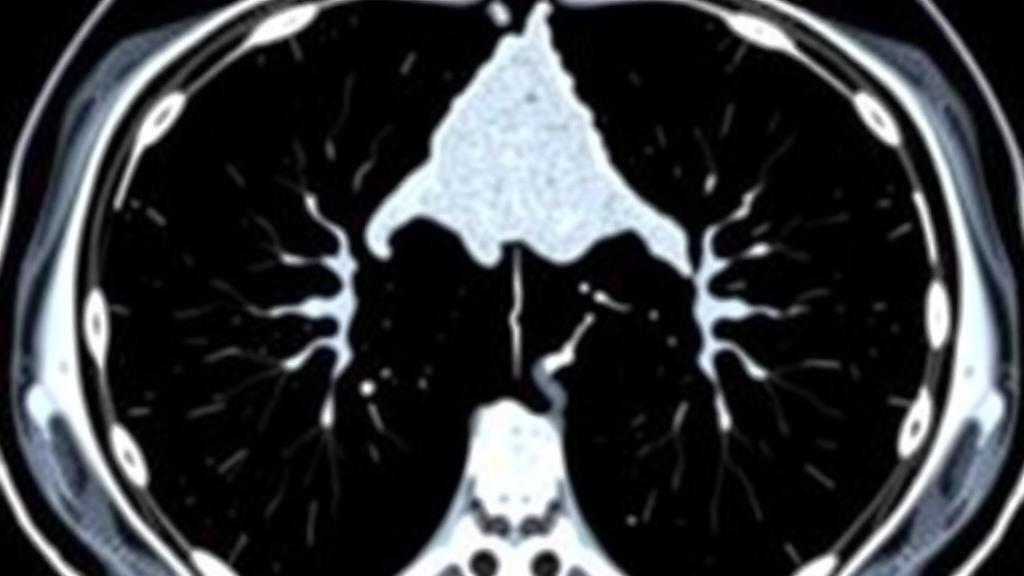
A routine computed tomography scan performed to stage a tumor may hold one of the most powerful clues to whether a patient with esophageal cancer will survive the grueling journey of modern treatment. A multicenter study published in the Journal of Cachexia, Sarcopenia and Muscle reports that the amount of skeletal muscle visible on a single cross-sectional image at the third lumbar vertebra, a measurement known as the skeletal muscle index, strongly predicts how well patients with locally advanced esophageal squamous cell carcinoma tolerate neoadjuvant immunochemotherapy, whether they make it to surgery, and how long they ultimately live. The findings, drawn from 461 patients treated at four Chinese hospitals between 2020 and 2023, suggest that muscle quantity, invisible to the bathroom scale, may be a modifiable determinant of success in one of oncology’s most demanding treatment pathways.
Neoadjuvant immunochemotherapy, in which a programmed cell death protein 1 inhibitor is combined with platinum-based chemotherapy before surgery, has transformed the outlook for locally advanced esophageal squamous cell carcinoma, the dominant histological subtype in China. Prospective trials such as ESCORT-Neo have demonstrated significantly improved pathological response and long-term survival. Yet the regimen exacts a heavy physiological toll. Even in highly selected trial cohorts, between 10.8 and 13.6 percent of patients never undergo surgical resection, with treatment-related adverse events and clinical intolerability contributing substantially to that attrition. Real-world evidence points to even harsher numbers, with severe toxicity affecting roughly 25 to 30 percent of patients and delaying or derailing planned operations. Because most prior analyses restricted themselves to patients who ultimately reached the operating table, early intolerance has remained a blind spot in the literature.
The research team, led from Fujian Medical University Union Hospital, addressed this gap by following every patient who initiated neoadjuvant therapy, not merely those who completed it. They introduced a novel framework called the neoadjuvant treatment tolerance grade, or NAT-TG, which integrates treatment intensity, dose reduction, delays, early discontinuation and completion of curative surgery into a single ordinal measure. Patients achieving full-intensity therapy followed by surgery were classified as NAT-TG1; those requiring modification but still reaching surgery were NAT-TG2; and those who failed to undergo curative surgery after starting treatment were NAT-TG3. This composite endpoint captures the entire neoadjuvant-to-surgery pathway, distinguishing physiological reserve from treatment feasibility in a way that conventional toxicity reporting does not.
The technical foundation of the study lies in imaging-based body composition analysis. Two trained radiologists, blinded to all clinical information and outcomes, independently selected an axial computed tomography image at the mid-pedicle level of the third lumbar vertebra, a standard landmark where skeletal muscle cross-sectional area correlates reliably with whole-body muscle mass. Using dedicated software, they segmented muscle tissue within attenuation thresholds of minus 29 to plus 150 Hounsfield units, manually correcting boundaries in roughly half of the cases to exclude misclassified adjacent structures. The resulting skeletal muscle area was normalized to height squared to yield the skeletal muscle index. Interobserver agreement was exceptional, with intraclass correlation coefficients of 0.994 for muscle area and 0.992 for the index, underscoring the reproducibility of the technique.
Rather than importing fixed cutoff values derived from Western populations, the investigators used X-tile software to derive sex-specific thresholds from their own cohort, defining low skeletal muscle index as less than 51.5 square centimeters per square meter in men and less than 37.5 in women. By this definition, 121 patients, or 26.2 percent of the cohort, had low muscle mass. Critically, most of these patients had a normal or even elevated body mass index, a phenomenon known as sarcopenic obesity or hidden muscle wasting. Body mass index, the researchers emphasize, cannot distinguish adipose tissue from lean mass, and patients with depleted muscle reserves hidden within an apparently adequate weight are precisely those most vulnerable to dose-limiting toxicity and premature treatment discontinuation.
The clinical consequences were stark. Only 48.8 percent of patients with low skeletal muscle index achieved the full-intensity treatment-and-surgery pathway, compared with 72.9 percent of those with preserved muscle. Unplanned failure to undergo curative surgery occurred in 20.7 percent of the low-muscle group versus 9.4 percent of the rest. Among the 57 patients who never reached the operating table, severe treatment-related toxicity was the dominant reason in 30 cases, persistent clinical or functional deterioration in 21, and disease progression in only six. Even after excluding the progression-related cases, low muscle mass remained independently associated with unfavorable tolerance, with an adjusted odds ratio of 2.51. Severe adverse events, particularly hematologic and gastrointestinal toxicities, were also markedly more frequent in the muscle-depleted group, affecting 36.4 percent versus 22.9 percent of patients.
Muscle depletion cast a long shadow beyond the neoadjuvant period. Among the 404 patients who underwent esophagectomy, those with low skeletal muscle index suffered severe postoperative complications, graded Clavien-Dindo III or higher, at nearly double the rate of their counterparts, 22.9 percent versus 11.0 percent, with pneumonia and pleural effusion driving much of the difference. After adjustment for age, sex, body mass index, serum albumin, operative approach, lymphadenectomy extent, tumor location and participating center, low muscle mass carried an adjusted odds ratio of 2.92 for severe surgical complications. Plausible mechanisms include reduced respiratory muscle strength, impaired cough effectiveness, altered drug distribution and clearance that increases systemic exposure to cytotoxic agents, and the chronic inflammation, metabolic dysregulation and immunosenescence that accompany muscle wasting, although the observational design cannot confirm direct causation.
Survival followed the same gradient. Over a median follow-up of 43 months, during which 165 deaths occurred, patients with low skeletal muscle index had a median overall survival of 35.0 months, while median survival had not been reached in the preserved-muscle group. Three-year overall survival was 49.4 percent with low muscle mass versus 74.4 percent without, and the association persisted across three progressively adjusted Cox models, with hazard ratios ranging from 2.17 to 2.38 in the intention-to-treat population. In the surgical cohort, low muscle mass independently predicted both poorer overall survival, with an adjusted hazard ratio of 1.91, and poorer disease-free survival, at 1.57. Bootstrap internal validation yielded optimism-corrected C-indices of approximately 0.68, indicating reasonably stable but moderate discrimination, and sensitivity analyses using published Prado and Fearon thresholds generally preserved the direction of the findings.
Perhaps the most provocative result emerged from an exploratory analysis of postoperative adjuvant immunotherapy. Among patients with low skeletal muscle index, those who received adjuvant immunotherapy within three months of surgery had substantially better three-year overall survival, 63.8 percent versus 43.1 percent, and disease-free survival, 59.0 percent versus 34.5 percent, compared with observation. The association survived multivariable adjustment, with adjusted hazard ratios of 0.47 for both death and recurrence or death, although a six-month landmark analysis designed to reduce immortal time bias produced estimates that crossed the null. No clear benefit was seen in patients with preserved muscle mass. The authors speculate that because muscle-depleted patients more often received modified or incomplete neoadjuvant therapy, they may carry greater residual disease burden, which postoperative immunotherapy could partially offset. They stress that this hypothesis-generating finding requires prospective validation.
The study has acknowledged limitations, including its retrospective design, potential residual confounding, cohort-derived cutoffs awaiting external validation, and the absence of muscle strength or physical performance measures, meaning the researchers assessed muscle quantity rather than clinically diagnosed sarcopenia. Nevertheless, the implications are considerable. The skeletal muscle index can be calculated from staging scans that esophageal cancer patients already undergo, at no additional cost or radiation exposure, offering a practical adjunct for baseline risk stratification. As immune checkpoint inhibitors and intensive multimodal regimens push treatment boundaries ever further, the study adds to mounting evidence that the body’s muscle compartment is not a passive bystander but an active determinant of who can endure, complete and benefit from modern cancer therapy, and that identifying hidden muscle depletion before treatment begins may open a window for nutritional and physical intervention.
Subject of Research: Skeletal muscle index as a predictor of tolerance and survival in neoadjuvant immunochemotherapy for oesophageal squamous cell carcinoma
Article Title: Low Skeletal Muscle Index Predicts Neoadjuvant Immunochemotherapy Tolerance and Survival in Oesophageal Squamous Cell Carcinoma
Article References: Low Skeletal Muscle Index Predicts Neoadjuvant Immunochemotherapy Tolerance and Survival in Oesophageal Squamous Cell Carcinoma. (n.d.). https://doi.org/10.1002/jcsm.70392
Image Credits: AI Generated
DOI: 10.1002/jcsm.70392
Keywords: skeletal muscle index, sarcopenia, oesophageal squamous cell carcinoma, neoadjuvant immunochemotherapy, body composition, PD-1 inhibitors, esophagectomy, treatment tolerance, postoperative complications, overall survival, CT imaging, adjuvant immunotherapy
Cite Scienmag News
Nathaniel Bowman. (October 3, 2026). Hidden Muscle Loss on CT Scans Predicts Who Tolerates Esophageal Cancer Immunotherapy. Scienmag. https://scienmag.com/hidden-muscle-loss-on-ct-scans-predicts-who-tolerates-esophageal-cancer-immunotherapy/
Nathaniel Bowman. "Hidden Muscle Loss on CT Scans Predicts Who Tolerates Esophageal Cancer Immunotherapy." Scienmag, 3 October 2026, https://scienmag.com/hidden-muscle-loss-on-ct-scans-predicts-who-tolerates-esophageal-cancer-immunotherapy/. Accessed 3 October 2026.
Nathaniel Bowman. "Hidden Muscle Loss on CT Scans Predicts Who Tolerates Esophageal Cancer Immunotherapy." Scienmag. October 3, 2026. https://scienmag.com/hidden-muscle-loss-on-ct-scans-predicts-who-tolerates-esophageal-cancer-immunotherapy/


