


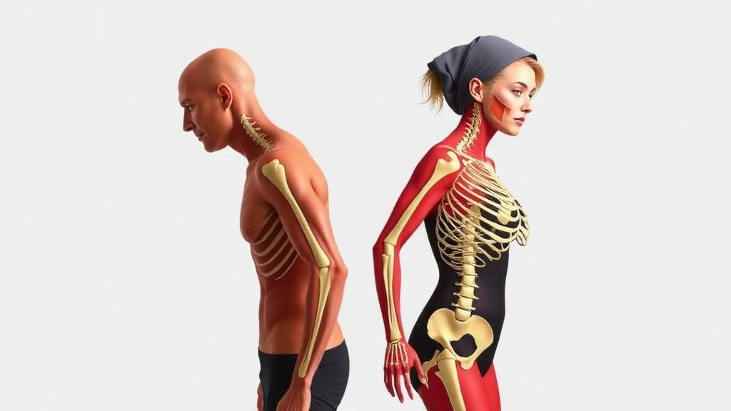
The three tissues that carry us through life—bone, muscle, and fat—do not age on a single schedule, and they certainly do not age the same way in men and women. That is the central message of a large cross-sectional study published in GeroScience, which mapped the trajectories of body composition across seven decades of adulthood in 9,717 Caucasian adults aged 20 to 90. Led by Jasminka Z. Ilich of Florida State University’s Institute for Successful Longevity, the research team used a nonionizing bioelectrical impedance technique called BIA-ACC to estimate total body bone mass, skeletal muscle mass, fat mass, and intramuscular adipose tissue, the fat that infiltrates muscle itself. By applying a flexible statistical method that makes no assumptions about the shape of aging curves, the investigators were able to pinpoint where those curves bend—and the results reveal strikingly divergent timetables for the two sexes, with potential consequences for how and when clinicians should screen for a newly emphasized condition known as osteosarcopenic adiposity.
Osteosarcopenic adiposity, abbreviated OSA, describes the coexistence of three normally separate problems: reduced bone density (osteopenia or osteoporosis), age-related loss of muscle mass and strength (sarcopenia), and excess or abnormally redistributed body fat. The concept matters because these tissues are not independent players. Bone, muscle, and fat communicate through shared hormonal, inflammatory, and mechanical pathways, and deterioration in one can accelerate decline in the others. Fat that invades muscle and even bone marrow is increasingly recognized not as a passive storage depot but as an active secretory organ that promotes chronic low-grade inflammation, insulin resistance, and catabolic signaling. When all three tissues fail together, the result is a vicious cycle of fragility, falls, fractures, and metabolic disease. Yet despite growing interest in the phenotype, the timing of its emergence—and whether it looks different in men and women—has remained poorly defined.
To address that gap, the team turned to LOESS, or locally estimated scatterplot smoothing, a nonparametric regression technique that fits flexible curves to data without imposing a predefined mathematical relationship between age and each body composition outcome. This is a crucial methodological choice. Traditional analyses often assume linear or simple polynomial declines, which can mask the true inflection points where tissue loss accelerates or decelerates. By letting the data speak, LOESS revealed the actual bends in the aging curves. The researchers also conducted sensitivity analyses, refitting all curves with varying stiffness parameters and computing confidence intervals, to ensure the identified inflection points were robust rather than artifacts of a particular smoothing setting. Prespecified operational thresholds for BIA-ACC-derived bone and muscle mass scores, along with sex-specific fat mass percentages, were used to classify participants as having reduced bone mass, reduced muscle mass, adiposity, or the combined OSA phenotype.
The sex differences were unmistakable. Men reached their peaks in bone and muscle mass earlier in life and then enjoyed a period of relative stability through midlife, before declining to lower values in later life than women of the same age. Women, by contrast, started adulthood with lower baseline bone and muscle values and displayed multiple inflection points early in life, suggesting a more turbulent developmental and early-adult trajectory. The most dramatic feature of the female curves was a sharp midlife downturn, particularly around the time of menopause—a pattern consistent with decades of research linking the loss of estrogen to accelerated bone resorption and adverse changes in muscle and fat distribution. Estrogen’s protective effects on bone remodeling and musculoskeletal performance are well documented, and the new curves provide a population-level visualization of exactly when that protection appears to be withdrawn.
Fat told a different story in each sex. In men, fat mass increased steadily and almost monotonically across adulthood, a slow and relentless accumulation with no dramatic inflection points. In women, fat mass followed multiphasic patterns, rising and falling in distinct phases across the life course, likely reflecting the interplay of reproductive hormones, childbearing years, and the menopausal transition. Intramuscular adipose tissue, however, behaved more uniformly: it increased progressively with age in both men and women, making fatty infiltration of muscle one of the most consistent markers of musculoskeletal aging in the dataset. This finding carries clinical weight, because IMAT is associated with impaired muscle quality, reduced mobility, and metabolic dysfunction, and it may represent an early warning sign that precedes overt losses of bone and muscle mass.
Perhaps the most sobering result concerned participants who met the operational BIA-ACC criteria for the OSA phenotype. These individuals were identified even among the youngest participants in the cohort, indicating that the triad of low bone mass, low muscle mass, and excess adiposity is not exclusively a disease of old age. Adults meeting the OSA criteria demonstrated less stable body composition trajectories overall, with earlier downturns in their bone and muscle components and more complex patterns in fat and intramuscular fat. In other words, the phenotype appears to mark a subgroup whose tissues begin declining ahead of schedule, compounding one another’s deterioration decades before the typical fragility window. The authors suggest that early identification of this phenotype could support preventive assessment of accelerated musculoskeletal decline and rising adiposity, potentially opening a window for intervention long before fractures or disability occur.
The study’s cross-sectional design warrants careful interpretation. Because each participant was measured at a single point in time, the curves represent differences between age groups rather than changes tracked within the same individuals over decades. Cohort effects—differences in nutrition, physical activity, and medical care between generations—could contribute to some of the observed patterns. Longitudinal studies, such as the Health, Aging and Body Composition Study cited by the authors, have documented long-term rates of change in musculoskeletal aging, and future work combining longitudinal follow-up with the inflection-point approach used here would strengthen causal inference. The cohort was also limited to Caucasian adults, and body composition norms and OSA prevalence may differ across ancestry groups, so generalization to other populations requires caution. Additionally, BIA-ACC, while nonionizing and suitable for very large samples, estimates rather than directly images tissue compartments; dual-energy X-ray absorptiometry and quantitative CT remain reference standards for bone and fat depots, and prior comparative work has examined bioimpedance against DEXA in postmenopausal women.
Even with those caveats, the scale and granularity of the analysis make it a valuable contribution to the biology of aging. The findings align with a broader shift in gerontology away from viewing aging as a smooth, uniform process and toward recognizing nonlinear dynamics, with molecular studies now identifying waves of change at specific ages across multiple organ systems. The menopause-related inflection in women’s bone and muscle curves echoes established clinical knowledge about postmenopausal osteoporosis, but the demonstration that men’s later-life values fall below women’s—despite their earlier, higher peaks—adds nuance to the conventional narrative that women are uniformly more vulnerable to musculoskeletal decline. It also underscores that men’s steady fat gain and progressive muscle fat infiltration deserve attention in their own right, particularly given the links between adipose tissue dysfunction, insulin resistance, and chronic disease.
For clinicians and the public, the practical takeaway is that the timing of risk is sex-specific and measurable. Women’s preventive efforts around bone and muscle may need to intensify in the years surrounding menopause, when the curves bend most sharply, while men’s risk profile may accumulate more insidiously through midlife fat gain and later-life tissue loss. Screening tools that can flag the OSA phenotype early—ideally nonionizing, inexpensive, and scalable, as bioimpedance-based approaches aim to be—could help identify people whose trajectories are already diverging from healthy aging. As the authors conclude, distinct sex-specific patterns exist, and the adverse association between the OSA phenotype and body composition trajectories suggests that catching this combined deterioration early may be one of the most effective strategies for preserving mobility, preventing fractures, and extending healthspan across the second half of life.
Subject of Research: Sex- and age-specific changes in bone, muscle, and fat mass across adulthood and their relation to the osteosarcopenic adiposity phenotype
Article Title: Sex- and age-specific patterns in bone, muscle, and fat across adulthood and their relation to osteosarcopenic adiposity phenotype: a cross-sectional study
Article References: Ilich, J. Z., Mills, J., Cvijetic, S., Barlow, E. M., Galijasevic, S., Boschiero, D., & Harman, J. (2026). Sex- and age-specific patterns in bone, muscle, and fat across adulthood and their relation to osteosarcopenic adiposity phenotype: a cross-sectional study. GeroScience. https://doi.org/10.1007/s11357-026-02496-1
Image Credits: AI Generated
DOI: 10.1007/s11357-026-02496-1
Keywords: aging, body composition, osteosarcopenic adiposity, sarcopenia, osteoporosis, intramuscular fat, menopause, bioelectrical impedance, LOESS regression, sex differences, bone health, adiposity
Cite Scienmag News
Beatrice Stafford. (September 30, 2026). Bone, Muscle, and Fat Follow Sharply Different Aging Timetables in Men and Women, Study of Nearly 10,000 Adults Finds. Scienmag. https://scienmag.com/bone-muscle-and-fat-follow-sharply-different-aging-timetables-in-men-and-women-study-of-nearly-10000-adults-finds/
Beatrice Stafford. "Bone, Muscle, and Fat Follow Sharply Different Aging Timetables in Men and Women, Study of Nearly 10,000 Adults Finds." Scienmag, 30 September 2026, https://scienmag.com/bone-muscle-and-fat-follow-sharply-different-aging-timetables-in-men-and-women-study-of-nearly-10000-adults-finds/. Accessed 30 September 2026.
Beatrice Stafford. "Bone, Muscle, and Fat Follow Sharply Different Aging Timetables in Men and Women, Study of Nearly 10,000 Adults Finds." Scienmag. September 30, 2026. https://scienmag.com/bone-muscle-and-fat-follow-sharply-different-aging-timetables-in-men-and-women-study-of-nearly-10000-adults-finds/


