


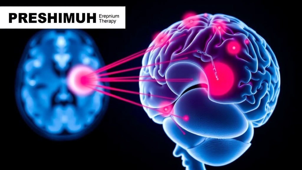
A rare and stubbornly difficult category of brain tumors has long forced adult patients and their physicians into an uncomfortable trade-off: the tumor itself is slow-growing and often survivable for decades, but the treatments available when surgery fails can inflict lasting damage on the very brain tissue that makes life worth living. A new study from Heidelberg University Hospital, published in the Journal of Neuro-Oncology, offers the most detailed look yet at how proton radiotherapy performs in this exact scenario, and the results are strikingly encouraging. In fourteen adults with progressive or surgically unfavorable pilocytic astrocytoma, proton beam therapy produced durable tumor control, marked radiographic shrinkage, and a toxicity profile that was overwhelmingly mild.
Pilocytic astrocytoma is a WHO grade 1 glioma, a tumor class that behaves far more indolently than the aggressive glioblastomas that dominate public awareness of brain cancer. These tumors are driven largely by alterations in the MAPK signaling pathway and, in children, are often cured by surgery alone. In adults, however, the disease is less well characterized. Adult tumors tend to arise in different locations, may be less amenable to complete resection, and can recur years or even decades after an initial operation. When they sit in the brainstem, the optic pathway, the hypothalamus, or deep midline ventricular structures, the risk of repeated surgery becomes unacceptable, leaving radiotherapy as the principal local option.
The Heidelberg team, led by Fabian J. K. Allmendinger and colleagues, assembled a retrospective cohort of fourteen adults treated with intensity-modulated proton radiotherapy between 2011 and 2025. The patients were young, with a median age of 23.5 years at treatment, and their tumors occupied some of the most anatomically treacherous territory in the brain: the cerebellum, the frontal and temporal lobes, the third and fourth ventricles, the suprasellar compartment, the mesencephalon, and the pineal region. Ten of the fourteen had undergone prior surgery, and in nine of those cases the operation had achieved only subtotal removal. For ten patients, proton therapy was salvage treatment for recurrent disease; for four, it was the primary local therapy when surgery was judged too risky.
The technical premise of proton therapy is elegantly simple. Unlike conventional photon beams, which deposit radiation along their entire path through tissue and exit on the far side of the target, protons carry a finite range. They release most of their energy in a sharply defined burst, the Bragg peak, at a depth determined by their initial energy, and deliver essentially nothing beyond it. By scanning pencil-thin proton beams across the tumor volume, clinicians can conform the dose to irregular targets while dramatically reducing the radiation bath received by uninvolved brain, the temporal lobes, the hippocampi, the optic apparatus, the hypothalamic-pituitary axis, and cerebral blood vessels. For patients expected to live for decades, that reduction in integral dose is not a cosmetic advantage; it is the difference between preserving and progressively eroding neurocognitive, endocrine, and visual function over a lifetime.
The prescribed dose in the cohort was a median of 54.0 Gy(RBE), delivered in roughly 29 fractions, with planning that co-registered treatment CT scans with contrast-enhanced T1-weighted and T2/FLAIR MRI and applied QUANTEC-based constraints for organs at risk. What followed was, by the standards of brain tumor therapy, remarkable. At last follow-up, bidirectional T2-weighted tumor measurements showed complete regression in three patients, partial response of at least fifty percent in seven, and a minor response in two. Only two patients experienced progressive disease. Perhaps most intriguingly, shrinkage frequently continued beyond the twelve-month landmark: ten of the twelve non-progressive patients kept improving after the standardized one-year assessment, a pattern consistent with the slow growth kinetics of grade 1 tumors and a warning against prematurely declaring treatment failure.
Survival outcomes matched the radiographic enthusiasm. Estimated overall survival was one hundred percent at both three and five years, with no deaths recorded within the first five years after therapy. Progression-free survival stood at 83.3 percent at both timepoints, and most progression events occurred early, meaning that patients who remained disease-free in the initial years tended to stay that way. Median clinical follow-up extended to nearly ten years, with vital status confirmed through national registry data, lending unusual depth to the survival analysis for a cohort of this size.
Toxicity was predominantly low grade. During treatment, the most common acute effects were focal alopecia in 64 percent of patients, fatigue and headache in 43 percent each, and radiodermatitis in 29 percent, and nearly all of these resolved in follow-up. Alopecia and skin reactions vanished entirely beyond the first months. Fatigue was the most persistent complaint, affecting three patients across all follow-up intervals, while mild cognitive impairment appeared in two. One patient with pre-existing postoperative visual deficits developed late grade 1 visual impairment, though optic nerve dose constraints had been met and imaging showed no optic neuropathy. Critically, no grade 4 or grade 5 events occurred at any point.
The study also shines a light on a subtle diagnostic trap known as radiation-induced contrast enhancement, or RICE. Within months to years after proton therapy, new areas of contrast uptake can appear on MRI within or near the irradiated volume, mimicking tumor progression on a scan. In this cohort, three patients developed RICE, yielding a three- and five-year RICE-free probability of 76.2 percent, with all events occurring in the early post-treatment period. One patient was initially misclassified as having progressed and was started on temozolomide chemotherapy, which had to be abandoned after a single cycle due to blood toxicity; retrospective review showed the enhancing lesion was radiation injury, not tumor, and bevacizumab sufficed. The authors emphasize that distinguishing RICE from true progression, using imaging morphology, temporal evolution, spatial relation to the dose distribution, and clinical course, is essential to avoid unnecessary chemotherapy in patients who otherwise face decades of life.
The findings arrive amid an evolving treatment landscape. Current EANO-EURACAN-SNO guidelines recommend maximal safe resection when feasible, with molecular testing for BRAF and MAPK alterations, and acknowledge reoperation, radiotherapy, and targeted agents such as BRAF and MEK inhibitors as options for recurrent or unresectable tumors. Recent multicenter data show chemotherapy with temozolomide achieves a median progression-free survival of only about twenty months in adult pilocytic astrocytoma, a figure that looks modest against the durable control seen here. The Heidelberg authors are careful, however, not to overreach: the study was retrospective, involved only fourteen patients, lacked a photon comparator, and carried incomplete molecular annotation, so it cannot prove that protons outperform photons, only that they deliver excellent control with acceptable morbidity in a selected, anatomically complex population.
Even with those caveats, the message for patients and clinicians is consequential. For adults facing a progressive pilocytic astrocytoma that surgery cannot safely reach, proton radiotherapy now has dedicated, adult-specific evidence behind it: near-universal radiographic response, complete five-year survival, and side effects that were mostly transient and mild. Because prospective trials are unlikely in a disease this rare, single-center cohorts like this one carry disproportionate weight. The study also reinforces a broader principle of modern radiation oncology, that in long-surviving patients with benign-behaving tumors, the true measure of a treatment is not only whether it controls the disease, but what quality of brain, and of life, it leaves behind ten, twenty, and thirty years later.
Subject of Research: Proton radiotherapy for progressive or surgically unfavorable adult pilocytic astrocytoma
Article Title: Efficacy and safety of proton radiotherapy for progressive or surgically unfavorable adult pilocytic astrocytoma: a single-center experience
Article References: Allmendinger, F. J. K., Cherniienko, Y., Lischalk, J., Walter, J., Mose, L., Wickert, R., Deng, M., Regnery, S., Wessel, L., Kozyra, K., Tessonnier, T., Krieg, S., Debus, J., König, L., & Adena-Eichkorn, T. (2026). Efficacy and safety of proton radiotherapy for progressive or surgically unfavorable adult pilocytic astrocytoma: a single-center experience. Journal of Neuro-Oncology, 179(3), Article 89. https://doi.org/10.1007/s11060-026-05798-8
Image Credits: AI Generated
DOI: 10.1007/s11060-026-05798-8
Keywords: proton radiotherapy, pilocytic astrocytoma, adult glioma, radiation oncology, RICE, tumor control, low-grade glioma, Bragg peak, neuro-oncology, radiation toxicity, MAPK pathway, brain tumors
Cite Scienmag News
Nathaniel Bowman. (September 27, 2026). Proton Beam Therapy Shows Durable Tumor Control in Rare Adult Brain Tumors. Scienmag. https://scienmag.com/proton-beam-therapy-shows-durable-tumor-control-in-rare-adult-brain-tumors/
Nathaniel Bowman. "Proton Beam Therapy Shows Durable Tumor Control in Rare Adult Brain Tumors." Scienmag, 27 September 2026, https://scienmag.com/proton-beam-therapy-shows-durable-tumor-control-in-rare-adult-brain-tumors/. Accessed 27 September 2026.
Nathaniel Bowman. "Proton Beam Therapy Shows Durable Tumor Control in Rare Adult Brain Tumors." Scienmag. September 27, 2026. https://scienmag.com/proton-beam-therapy-shows-durable-tumor-control-in-rare-adult-brain-tumors/


