


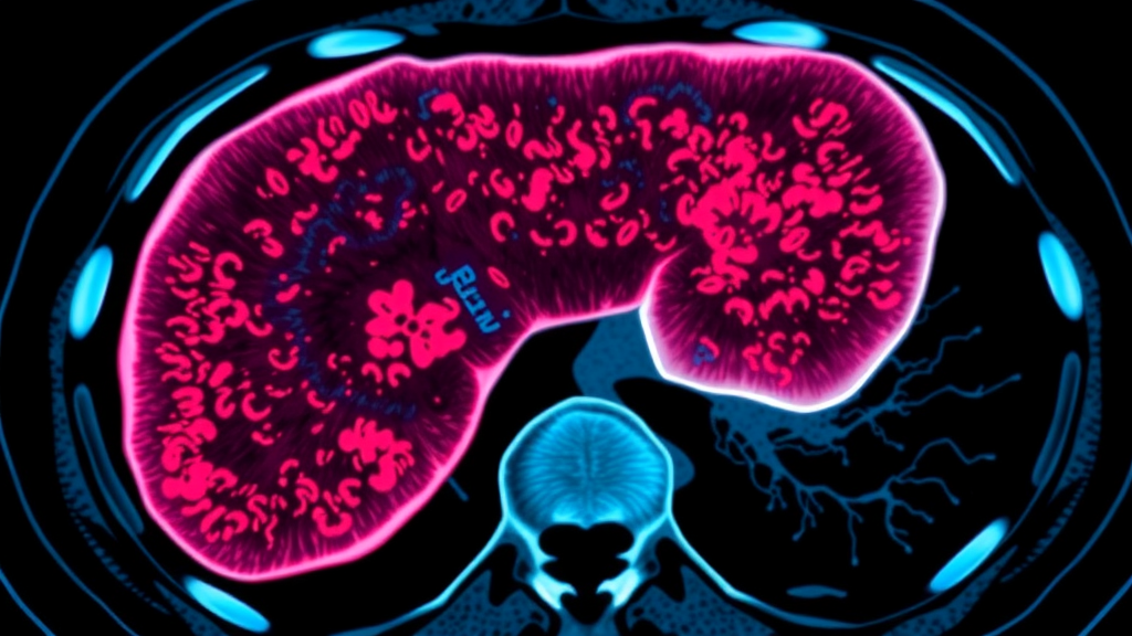
A routine CT scan may soon do more than show the size and location of a liver tumor. A large multicenter study published in BMC Medical Imaging reports that a computed tomography-based artificial intelligence framework can predict, noninvasively, whether a hepatocellular carcinoma belongs to an aggressive molecular subtype defined by the expression of a protein called cytokeratin 19, or CK19. The finding matters because CK19 status, which today can only be established after surgical removal of the tumor and laboratory staining of the tissue, is strongly linked to invasive behavior, early recurrence, and poor outcomes in liver cancer patients.
Hepatocellular carcinoma is the most common form of primary liver cancer and a leading cause of cancer deaths worldwide. Not all hepatocellular carcinomas behave the same way. A substantial fraction, roughly forty to fifty percent in surgical series, express CK19, an intermediate filament protein normally found in the bile ducts rather than in mature liver cells. When hepatocytes, the main working cells of the liver, re-express or retain this protein, the tumor tends to behave more aggressively: it invades surrounding tissue more readily, metastasizes earlier, and responds less favorably to standard treatments. Clinicians would therefore like to know a patient’s CK19 status before committing to a treatment plan, but under current practice that information arrives only after pathology has examined the resected specimen.
The research team, led by Yongming Tan, Siya Xu, and Shucheng Yang of the First Affiliated Hospital of Nanchang University, together with colleagues across three institutions in Jiangxi Province, China, set out to close this gap with imaging. Their retrospective analysis included 1,029 patients with surgically treated hepatocellular carcinoma drawn from three hospitals: the First Affiliated Hospital of Nanchang University, Jiangxi Cancer Hospital, and the Second Affiliated Hospital of Nanchang University. For each patient, the investigators collected preoperative contrast-enhanced CT images, manually delineated tumor masks, clinicopathological characteristics, CK19 immunohistochemical results, and follow-up data. Of the 1,029 patients, 459, or 44.6 percent, proved CK19-positive, while 570, or 55.4 percent, were CK19-negative, providing a balanced foundation for model development and validation.
The technical core of the study is a method the authors call recursive nested habitat analysis. The approach builds on a concept known as habitat imaging, which treats a tumor not as a uniform mass but as a collection of spatially distinct subregions, or habitats, each with its own imaging signature. In tumors, these habitats often correspond to biologically meaningful zones such as viable proliferating tissue, necrotic cores, and regions near feeding blood vessels that differ in oxygen and nutrient supply. Conventional radiomics, which extracts quantitative features from imaging data, typically summarizes the whole tumor in aggregate. Habitat analysis instead models localized patterns, capturing the internal architecture that aggregate features smooth over.
What makes the new framework distinctive is its recursive, nested structure. Rather than segmenting the tumor into habitats once, the algorithm models spatially localized imaging patterns and then progressively refines the regions with the highest probability of carrying the molecular signal of interest. In practice, the analysis zooms into high-probability subregions, characterizes them, and aggregates the information into a single habitat score. The idea is analogous to how a pathologist might scan a slide at low magnification, identify suspicious areas, and then examine those areas at higher magnification. The study also demonstrates that the framework is not merely a black box: the authors report that their nested habitat score consistently outperformed lower-level habitat scores, indicating that the recursive refinement itself, and not just any subdivision of the tumor, carries the predictive information.
The results are notable for their external validation, a step many radiomics studies skip. The model achieved an area under the receiver operating characteristic curve, or AUC, of 0.808, with a 95 percent confidence interval of 0.760 to 0.854, in external validation cohorts, meaning it discriminated CK19-positive from CK19-negative tumors with moderate to good accuracy on data from institutions not used to build it. Adding the habitat score to standard clinical variables improved CK19 prediction compared with a clinical model alone. Prespecified ablation analyses, in which the researchers systematically varied patch scales, enrichment fractions, and aggregation strategies, showed that performance remained stable across these design choices, a sign that the signal is robust rather than an artifact of one particular configuration.
The study is equally candid about its limits. Although the team explored whether the habitat score could predict early recurrence and stratify patients by recurrence-free survival, the nested habitat score did not provide significant incremental improvement for early recurrence prediction, nor did it retain independent prognostic value for recurrence-free survival after external validation. The authors explicitly frame these recurrence analyses as secondary and exploratory, cautioning against overinterpreting them. This kind of negative result, reported transparently, strengthens confidence in the positive finding: the imaging signal genuinely tracks CK19 biology without overstating what it can do.
Clinically, the implications are substantial. If validated prospectively, a CT-based CK19 surrogate could influence decisions well before surgery. Patients whose imaging suggests CK19-positive disease might be prioritized for more aggressive surveillance, considered for adjuvant therapies, or guided toward treatment strategies that account for the subtype’s invasive behavior. The decision curves published with the study suggest potential net benefit over threshold probabilities between 0.10 and 0.70, although the authors stress that the threshold represents consideration of additional CK19-oriented assessment, not a validated treatment action. The framework also fits into a broader movement called radiopathomics, which seeks to connect large-scale imaging features with pathological and molecular ground truth, effectively teaching algorithms to see what only a microscope could previously reveal.
The methodological rigor underpinning the work deserves attention. The study underwent ethics approval under a formal reliance arrangement across the participating centers, analyzed only de-identified data, and included technical image-mask compatibility checks and feature-level inter-reader reproducibility assessments to verify that the extracted features were reliable. Calibration was evaluated center by center in external validation, and paired bootstrap comparisons quantified the incremental value of the nested approach over simpler alternatives. The work was funded by several Jiangxi Province research programs, and the authors declare no competing interests. The article is open access under a Creative Commons Attribution 4.0 license, and because it was shared early, the published version may be replaced by a final Version of Record after further edits.
What comes next is clear from the authors’ own conclusions. Before any clinical implementation, prospective studies integrating standardized imaging protocols, pathology, and multi-omics data will be required to confirm that the CT-derived habitat score holds up in real-world, forward-looking settings. Still, the study adds hepatocellular carcinoma to the growing list of cancers for which spatially aware, AI-driven imaging analysis can peer beneath the surface of a routine scan and infer molecular characteristics. For the hundreds of thousands of patients diagnosed with liver cancer each year, the prospect of learning a tumor’s molecular personality from images taken before a single incision is made represents a meaningful step toward precision oncology, delivered by the same contrast-enhanced CT that has been part of standard care for decades.
Subject of Research: Noninvasive prediction of CK19 expression in hepatocellular carcinoma using CT-based recursive nested habitat imaging analysis
Article Title: CT-based CK19-oriented recursive nested habitat analysis for noninvasive phenotyping of hepatocellular carcinoma: a multicenter external validation study
Article References: Tan, Y., Xu, S., Yang, S., Zhou, Y., Zhao, J., Wang, W., Liu, M., & Liu, J. (2026). CT-based CK19-oriented recursive nested habitat analysis for noninvasive phenotyping of hepatocellular carcinoma: a multicenter external validation study. BMC Medical Imaging. https://doi.org/10.1186/s12880-026-02821-2
Image Credits: AI Generated
DOI: 10.1186/s12880-026-02821-2
Keywords: hepatocellular carcinoma, cytokeratin 19, computed tomography, habitat imaging, radiomics, artificial intelligence, radiopathomics, molecular phenotyping, tumor heterogeneity, external validation, liver cancer, imaging biomarker
Cite Scienmag News
Nathaniel Bowman. (September 23, 2026). AI Reads Liver Tumors on CT Scans to Predict Aggressive Cancer Subtype Without Biopsy. Scienmag. https://scienmag.com/ai-reads-liver-tumors-on-ct-scans-to-predict-aggressive-cancer-subtype-without-biopsy/
Nathaniel Bowman. "AI Reads Liver Tumors on CT Scans to Predict Aggressive Cancer Subtype Without Biopsy." Scienmag, 23 September 2026, https://scienmag.com/ai-reads-liver-tumors-on-ct-scans-to-predict-aggressive-cancer-subtype-without-biopsy/. Accessed 23 September 2026.
Nathaniel Bowman. "AI Reads Liver Tumors on CT Scans to Predict Aggressive Cancer Subtype Without Biopsy." Scienmag. September 23, 2026. https://scienmag.com/ai-reads-liver-tumors-on-ct-scans-to-predict-aggressive-cancer-subtype-without-biopsy/
![Grilled and Smoked Food Carcinogen Benzo[a]pyrene Linked to Prostate Cancer Through Four Key Genes](https://scienmag.com/wp-content/plugins/wp-fastest-cache-premium/pro/images/blank.gif)

