


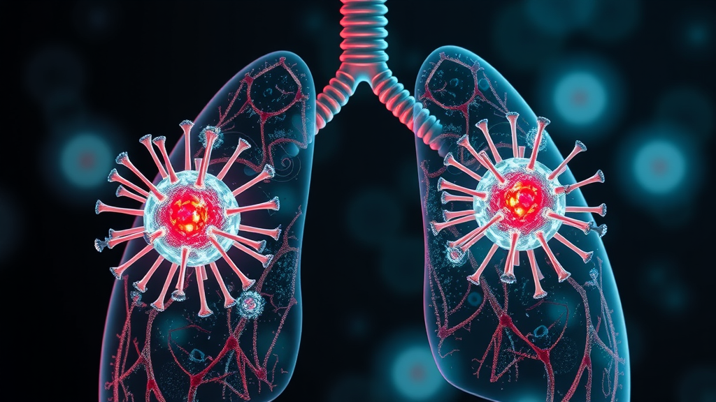
Congenital diaphragmatic hernia is one of the most serious structural birth defects encountered in neonatal medicine, and for decades its clinical course has been dominated by two intertwined problems: underdeveloped lungs and dangerously high blood pressure in the vessels of those lungs. New research now points to a contributor that has received far less attention than the anatomical defect itself. According to findings published in Pediatric Research, the endothelial cells that line the blood vessels of infants with congenital diaphragmatic hernia show measurable alterations in their bioenergetics, meaning the way these cells generate and manage energy appears fundamentally changed in the disease state. The observation reframes a condition long treated as primarily a mechanical and structural problem as one that also involves a deep cellular metabolic component, potentially opening new avenues for diagnosis and therapy.
The immediate consequence of congenital diaphragmatic hernia is structural. A hole in the diaphragm, the muscular partition separating the chest from the abdomen, allows abdominal organs such as the stomach, liver, and intestines to migrate into the chest cavity during fetal development. The crowding effect is severe: the developing lungs are compressed at precisely the window of gestation when their airways and vascular trees should be branching and expanding. The result is pulmonary hypoplasia, lungs that are smaller, simpler in architecture, and less able to support gas exchange after birth. But the mechanical compression is only part of the story. Newborns with this condition frequently develop persistent pulmonary hypertension, in which the resistance of the pulmonary circulation is so high that the heart struggles to pump blood through the lungs, leading to critical oxygen deprivation that can be fatal even with aggressive intervention.
The pulmonary hypertension associated with the condition has always hinted at a vascular problem, and vascular problems invite scrutiny of the endothelium. Endothelial cells form the inner lining of every blood vessel, and in the pulmonary circulation they do far more than serve as a passive barrier. They regulate vascular tone by producing nitric oxide, a potent vasodilator; they orchestrate angiogenesis, the growth of new vessels; they modulate inflammation and blood clotting; and they communicate constantly with the smooth muscle cells that wrap around vessels and control their caliber. In pulmonary hypertension of any origin, endothelial dysfunction is a recurring theme, and congenital diaphragmatic hernia is no exception. Previous work has described abnormal vascular remodeling in the lungs of affected infants, including medial thickening of pulmonary arteries and aberrant extension of muscle into distal, normally non-muscularized vessels. What the new study adds is a specific mechanistic suspect: disturbed energy metabolism within the endothelial cells themselves.
Bioenergetics refers to the set of biochemical pathways by which a cell converts nutrients into usable chemical energy, chiefly in the form of adenosine triphosphate, or ATP. For most cells in the body, the mitochondria are the power plants, and their efficiency determines whether a cell thrives or struggles under stress. Endothelial cells are unusual in this respect. Unlike many other cell types, healthy endothelial cells generate the majority of their ATP through glycolysis, the anaerobic breakdown of glucose, even when oxygen is abundantly available. This glycolytic preference is not a quirk; it is functionally important. It spares oxygen for the surrounding tissue, positions the endothelium to survive in low-oxygen environments during vessel sprouting, and produces biosynthetic intermediates needed for the rapid proliferation of new vessel lining during angiogenesis. Any disruption to this finely tuned metabolic balance can therefore ripple outward, impairing nitric oxide production, promoting oxidative stress, and destabilizing the vessel wall.
It is against this background that the reported alterations in endothelial bioenergetics take on significance. The study investigated how endothelial cells in the setting of congenital diaphragmatic hernia differ in their energy-generating machinery, examining markers of mitochondrial function and respiratory activity. Cells under metabolic stress typically exhibit a recognizable signature: diminished mitochondrial respiratory capacity, altered balance between oxidative phosphorylation and glycolysis, elevated production of reactive oxygen species, and reduced ability to adapt when energy demands spike. Each of these changes can feed a vicious cycle relevant to pulmonary hypertension. Mitochondrial dysfunction impairs the enzymes that synthesize nitric oxide, and when the endothelial nitric oxide synthase enzyme becomes uncoupled, it can actually generate superoxide instead of the protective vasodilator molecule. The resulting oxidative burden damages proteins, lipids, and DNA inside the vessel wall, encouraging the very remodeling and vasoconstriction that define hypertensive lung circulation.
The clinical stakes of this line of investigation are considerable. Infants with congenital diaphragmatic hernia are among the most intensively supported patients in neonatal intensive care, often requiring high-frequency ventilation, inhaled nitric oxide therapy, and in severe cases extracorporeal membrane oxygenation, a heart-lung bypass machine that takes over gas exchange while the infant’s own circulation stabilizes. Despite these measures, mortality remains substantial, particularly in cases diagnosed early with severe lung involvement. One of the most difficult problems clinicians face is predicting which infants will deteriorate and tailoring the intensity of support accordingly. If endothelial bioenergetic status proves to correlate with disease severity, metabolic markers could eventually supplement current predictors of outcome, giving intensive care teams a biochemical window into the state of the pulmonary vasculature that imaging and blood gas measurements cannot fully provide.
There is also a therapeutic dimension to consider. Metabolism has moved to the center of vascular biology research over the past two decades, and drugs that modulate mitochondrial function or cellular energy pathways already exist for other conditions. The concept of a metabolically targeted therapy for pulmonary vascular disease is no longer speculative in principle; several metabolic modulators, including agents that influence fatty acid oxidation and mitochondrial dynamics, are under investigation for pulmonary hypertension more broadly. If the endothelial energy deficit identified in congenital diaphragmatic hernia can be corrected, or even partially compensated, it could complement existing treatments that work through entirely different mechanisms. Inhaled nitric oxide, for example, acts downstream to relax vessels, but it does nothing to repair the underlying endothelial dysfunction that limits natural nitric oxide production. A therapy aimed at restoring mitochondrial health would attack the problem at its cellular source.
Careful interpretation remains essential. The connection between altered endothelial bioenergetics and clinical outcomes in this disease is an association that must now be interrogated further. Key questions include whether the metabolic changes observed are a cause of the pulmonary vascular pathology or a consequence of it, whether they are present before birth or emerge postnatally under the stress of intensive care, and whether they can be detected reliably in accessible clinical samples such as blood or in cells grown from patient-derived material. Animal models of the condition, including the nitrofen-induced rodent model widely used in the field, will likely play an important role in establishing causality, since they allow researchers to examine lung vessels at defined developmental stages that are difficult or impossible to access in human fetuses. The heterogeneity of the disease, which ranges from mild cases detected incidentally to lethal ones diagnosed prenatally, adds another layer of complexity to any attempt at generalization.
For the broader research community, the study sits at the intersection of two fields that have been converging rapidly: developmental vascular biology and cellular metabolism. The same bioenergetic principles that govern tumor angiogenesis and wound healing apply to the construction of the fetal pulmonary circulation, and perturbations of those principles during gestation may leave lasting functional fingerprints. Congenital diaphragmatic hernia, precisely because its vascular pathology is so pronounced and its clinical course so well documented, offers a natural setting in which to test whether endothelial energetics shape developmental outcomes. As follow-up work refines these findings, the hope is that a condition whose treatment has long been defined by supportive care will begin to yield to targeted, mechanism-based interventions, giving newborns with this devastating diagnosis a better chance at healthy, unrestricted breathing from their very first days.
Subject of Research: Endothelial cell bioenergetic alterations in congenital diaphragmatic hernia
Article Title: Alterations of endothelial cell bioenergetics in congenital diaphragmatic hernia
Article References: Emrick, B. F., Zhevlakova, I., Novotny, M., Mavrakis, L., Mulya, A., Cass, D. L., Miyasaka, E., Byzova, T. V., Asosingh, K., Erzurum, S. C., & Robertson, J. O. (2026). Alterations of endothelial cell bioenergetics in congenital diaphragmatic hernia. Pediatric Research. https://doi.org/10.1038/s41390-026-05447-w
Image Credits: AI Generated
DOI: 10.1038/s41390-026-05447-w
Keywords: congenital diaphragmatic hernia, endothelial cells, bioenergetics, mitochondria, pulmonary hypertension, pulmonary hypoplasia, nitric oxide, neonatal care, vascular remodeling, pediatric research, Alterations, endothelial
Cite Scienmag News
Harold Sullivan. (September 20, 2026). Cell Power Plants Emerge as New Suspects in Birth Defect That Cripples Lungs. Scienmag. https://scienmag.com/cell-power-plants-emerge-as-new-suspects-in-birth-defect-that-cripples-lungs/
Harold Sullivan. "Cell Power Plants Emerge as New Suspects in Birth Defect That Cripples Lungs." Scienmag, 20 September 2026, https://scienmag.com/cell-power-plants-emerge-as-new-suspects-in-birth-defect-that-cripples-lungs/. Accessed 20 September 2026.
Harold Sullivan. "Cell Power Plants Emerge as New Suspects in Birth Defect That Cripples Lungs." Scienmag. September 20, 2026. https://scienmag.com/cell-power-plants-emerge-as-new-suspects-in-birth-defect-that-cripples-lungs/


