


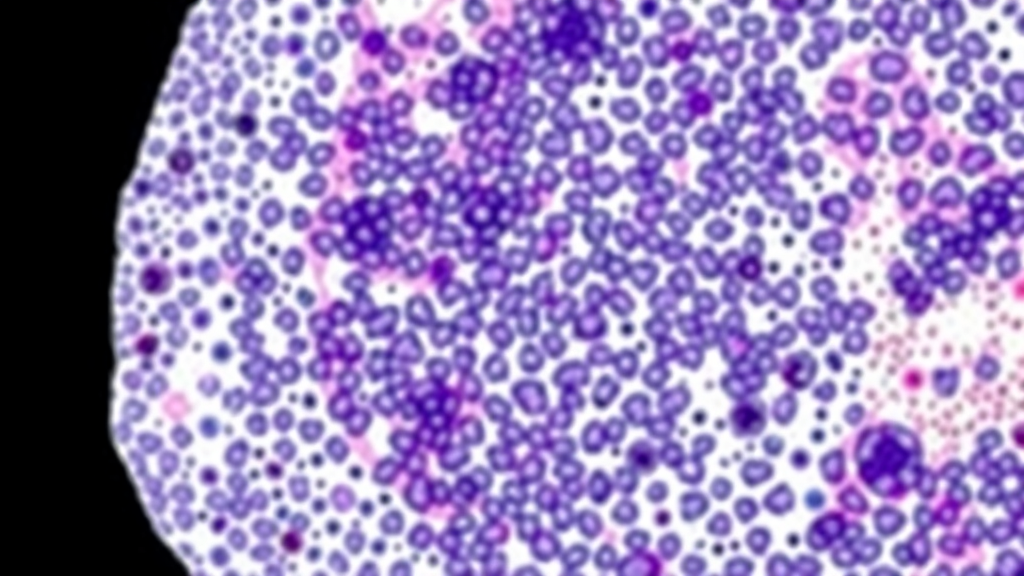
Tissue microarrays have quietly become one of the workhorses of modern cancer research, allowing hundreds of tissue cores from different patients to be packed onto a single microscope slide and scanned in one pass. But anyone who has worked with these dense grids of tissue knows the hidden labor involved: before the data can be analyzed, each individual core must be digitally sliced back out of the whole-slide image and reconnected to the correct patient metadata. A new open-source tool called TMA-Grid, described in BMC Bioinformatics, promises to make that painstaking process dramatically easier by running entirely inside a web browser, with no installation, no downloads, and no data ever leaving its storage location.
The researchers, led by Aaron Ge and colleagues at the National Cancer Institute’s Division of Cancer Epidemiology and Genetics, set out to solve a problem that has nagged the digital pathology community for years. Tissue microarrays are assembled by punching small cylindrical cores out of donor tissue blocks and arranging them in a precise grid on a recipient paraffin block. In practice, however, the assembly process, repeated sectioning, and high-resolution scanning introduce misalignments, missing cores, folds, and other artifacts. Any software that blindly assumes a perfect grid risks extracting the wrong tissue or mislabeling samples, errors that can silently corrupt downstream epidemiologic analyses.
Conventional de-arraying solutions have typically been desktop applications that must be downloaded, installed, and maintained, often on specific operating systems. They frequently offer limited interactivity, leaving users unable to correct the automated guesses the software makes when a core is shifted, absent, or duplicated. The NCI team argues that this rigidity has real consequences for large-scale studies, such as cancer epidemiology cohorts, where thousands of TMA slides must be processed reliably and where a single misassigned core can compromise the statistical integrity of a biomarker analysis.
TMA-Grid’s answer is a two-stage workflow that combines machine learning with human oversight. At its core is a convolutional neural network trained to segment tissue from background, detecting the individual cores on a scanned slide regardless of staining method or scanner idiosyncrasies. The network runs in the browser itself, powered by TensorFlow.js, which means the computational heavy lifting of tissue segmentation happens on the user’s own machine rather than on a remote server. This design choice has a welcome side effect for privacy-conscious institutions: sensitive patient slides never need to be uploaded anywhere to be processed.
Once cores are detected, the application applies an interactive grid-estimation algorithm that maps the detected tissue spots onto expected row and column positions, reconstructing the logical array layout that the pathologist originally designed. Crucially, this is not a black box. At every step, users can inspect, adjust, and override both the segmentation results and the grid assignment, adding missing cores, deleting artifacts, or nudging boundaries by hand. The developers describe this marriage of automated detection and user-driven correction as the central design principle: the machine does the tedious work, but the human retains final authority over what counts as a valid core.
The technical architecture follows what the authors call a zero-footprint approach. Nothing is installed on the user’s computer, and nothing is copied or cached to a server. The application operates on source images in place, whether they sit on a local hard drive, a remote institutional server, or cloud storage, reading them through standard web protocols. This means a researcher in Calgary, a statistician in London, and a bioinformatician in Maryland can all process slides from the same archive without ever duplicating multi-gigabyte whole-slide image files, a nontrivial benefit when studies involve hundreds of slides and limited bandwidth.
Beyond convenience, the team emphasizes adherence to FAIR principles, making the software Findable, Accessible, Interoperable, and Reusable. The application and all of its components are freely available and open source, and the authors designed its building blocks, from the segmentation model to the visualization layer built on the OpenSeadragon deep-zoom viewer, so that other groups can repurpose them in their own web-based digital pathology pipelines. In an era when reproducibility concerns shadow much of computational biology, publishing a fully open, browser-native tool with reusable components is a deliberate statement about how research software should be built and shared.
The practical implications reach well into cancer epidemiology. The work emerged from the NCI Intramural Research Program within the Division of Cancer Epidemiology and Genetics and its Episphere platform, an environment built for large-scale collaborative cancer research. Cohorts such as the Polish Breast Cancer Study and other population studies rely on tissue microarrays stained with hematoxylin and eosin or immunohistochemistry markers to screen candidate biomarkers across thousands of tumors. A reliable, interactive de-arraying tool that pathologists and technicians can use without IT support could compress what used to be a bottleneck into a routine step, accelerating the pace at which tissue-based discoveries translate into population-level insights.
The tool also reflects a broader shift in scientific software toward running where the data already live. As whole-slide imaging has exploded in scale, with single slides now routinely exceeding a gigapixel, moving files between workstations has become one of the biggest friction points in digital pathology. Zero-footprint web applications sidestep the problem entirely, and TMA-Grid demonstrates that even deep-learning-based image analysis, traditionally the domain of GPU-equipped servers, can now be pushed to the client side of the browser. The authors suggest the same pattern can generalize to other digital pathology tasks, from quality control to annotation workflows.
TMA-Grid is freely available as an open-source application, and its authors, drawn from the National Cancer Institute, the University of Calgary, Leidos Biomedical Research, and The Institute of Cancer Research in London, position it as both a practical solution for working TMA laboratories and a reusable foundation for the next generation of browser-based pathology tools. For a field whose progress increasingly depends on extracting maximum value from irreplaceable tissue collections, a tool that pairs automated intelligence with transparent human control may prove to be exactly what the pathologist ordered.
Subject of Research: Open-source browser-based tissue microarray de-arraying using deep learning for digital pathology
Article Title: TMA-Grid: an open-source, zero-footprint web application for FAIR tissue microarray de-arraying
Article References: Ge, A., Saha, M., Duggan, M. A., Lenz, P., Abubakar, M., García-Closas, M., Balasubramanian, J., Almeida, J. S., & Bhawsar, P. M. (2026). TMA-Grid: an open-source, zero-footprint web application for FAIR tissue microarray de-arraying. BMC Bioinformatics. https://doi.org/10.1186/s12859-026-06638-2
Image Credits: AI Generated
DOI: 10.1186/s12859-026-06638-2
Keywords: tissue microarray, de-arraying, digital pathology, whole slide imaging, FAIR software, web application, TensorFlow.js, OpenSeadragon, convolutional neural network, cancer epidemiology, open source, zero-footprint
Cite Scienmag News
Drew Townsend. (September 13, 2026). Browser-Based TMA-Grid Tool Brings FAIR, Zero-Footprint De-Arraying to Digital Pathology. Scienmag. https://scienmag.com/browser-based-tma-grid-tool-brings-fair-zero-footprint-de-arraying-to-digital-pathology/
Drew Townsend. "Browser-Based TMA-Grid Tool Brings FAIR, Zero-Footprint De-Arraying to Digital Pathology." Scienmag, 13 September 2026, https://scienmag.com/browser-based-tma-grid-tool-brings-fair-zero-footprint-de-arraying-to-digital-pathology/. Accessed 13 September 2026.
Drew Townsend. "Browser-Based TMA-Grid Tool Brings FAIR, Zero-Footprint De-Arraying to Digital Pathology." Scienmag. September 13, 2026. https://scienmag.com/browser-based-tma-grid-tool-brings-fair-zero-footprint-de-arraying-to-digital-pathology/


