


In a striking reminder that the humble blood smear still has life in it, laboratory physicians in Southwest China have reported the case of a three-year-old boy whose subcutaneous masses were traced to a parasitic worm infection only after a sharp-eyed technician spotted degranulated eosinophils on a peripheral blood film. The case, published in Acta Parasitologica, details how an unexpected microscopic clue led to a diagnosis of ectopic paragonimiasis, a food-borne parasitic disease that had eluded detection by conventional means.
Paragonimiasis is caused by trematode flatworms of the genus Paragonimus, lung flukes that typically infect humans who consume raw or undercooked freshwater crabs or crayfish harboring the parasite’s larval stage. Once ingested, the parasites excyst in the intestine, penetrate the intestinal wall, and migrate through the peritoneal cavity and diaphragm into the lungs, where they mature into adult flukes, encyst, and begin producing eggs. The classic presentation involves chronic cough, hemoptysis, and pulmonary cysts that can mimic tuberculosis or lung cancer. But the parasite is notorious for wandering off course. When juvenile worms fail to reach the lungs and instead settle in the brain, liver, skin, or subcutaneous tissue, the result is ectopic paragonimiasis, a form of the disease that is notoriously difficult to diagnose because neither eggs nor adult worms are found in sputum or stool.
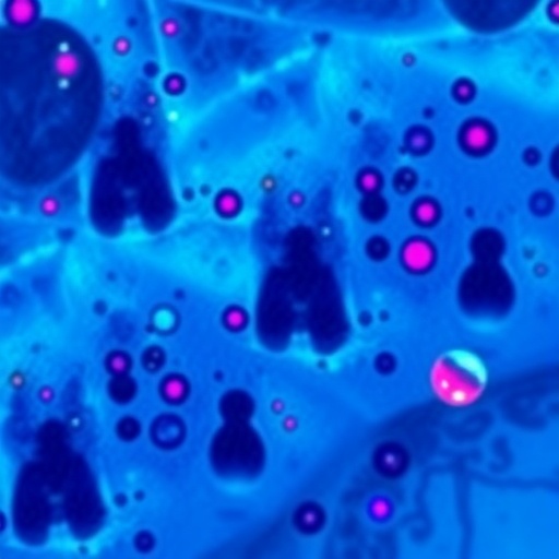
In this case, the young patient presented with subcutaneous masses, a clinical picture that could plausibly have been attributed to any number of far more common conditions in a small child, from benign lipomas to bacterial abscesses to malignant tumors. Without eggs detectable in biological samples and without adult parasites recoverable from the masses, the diagnostic team had little to go on. It was here that the peripheral blood smear provided the unexpected clue. Rather than the intact, crystalline granules that define healthy eosinophils, the smear revealed cells whose granules had been discharged, leaving pale, partially vacuolated cytoplasm. This phenomenon, known as degranulation or the appearance of hypogranular eosinophils, reflects intense activation of the eosinophil lineage, which occurs when the immune system is confronting a multicellular parasite.
Eosinophils are white blood cells armed with highly basic granules containing cytotoxic proteins such as major basic protein and eosinophil cationic protein. Their primary evolutionary purpose is defense against helminths, the parasitic worms that are too large to be phagocytosed. When eosinophils encounter parasitic antigens, they release the contents of these granules in an attempt to kill or damage the invader. The activated cells, having released their granules, appear under the microscope as degranulated or hypogranular forms. The presence of large numbers of these cells in circulation is a signal that the body is actively battling something worm-like, even if the parasite itself is hidden in tissue far from the blood.
What makes this case particularly instructive is the fact that the automated hematology analyzers widely used in modern hospitals failed to flag the significance of what was happening. These machines classify white blood cells based on physical properties such as size and internal complexity, and they struggle with abnormal cell morphology. Previous research has documented a mismatch between Sysmex XN-series analyzers and manual smear review, specifically in the context of hypogranular eosinophils being misclassified as platelets or other cell types. Hypogranular eosinophils have been described in eosinophilic gastrointestinal disease, but their association with parasitic infection remains underappreciated in an era of increasing automation. The authors of the current report emphasize that the visual examination of a peripheral blood smear remains an indispensable diagnostic tool, particularly when automated results do not align with the clinical picture or when an unexplained eosinophilia is present.
Building on the morphological suspicion generated by the smear, the diagnostic team pursued two complementary lines of investigation. First, they conducted an epidemiological assessment, recognizing that the boy lived in an area where Paragonimus is endemic and where consumption of raw or undercooked freshwater crustaceans is culturally common. Second, they ordered a serological test for Paragonimus-specific antibodies, which returned a positive result. The convergence of degranulated eosinophilia on the smear, compatible epidemiological exposure, and confirmatory serology established the diagnosis of ectopic paragonimiasis without the need for surgical excision of the mass or histopathological identification of parasite tissue.
The clinical significance of this approach extends well beyond a single pediatric case. Paragonimiasis is classified by the World Health Organization among the food-borne trematode infections of public health importance, with tens of millions of people at risk globally. Although China bears a substantial share of the global burden, with multiple Paragonimus species and a wide range of intermediate hosts documented across the country, the disease is increasingly recognized in nonendemic regions as well, driven by international travel, immigration, and the global food trade. Ectopic presentations, in which the parasite lodges in the skin, brain, liver, or orbit, are particularly treacherous diagnostically. Published case reports describe ectopic paragonimiasis mimicking hepatocellular carcinoma, orbital cellulitis, and even requiring surgical decortication when it presented as massive empyema in the chest. In one analysis of 685 cases of pleuropulmonary paragonimiasis diagnosed over 22 years, the breadth of radiological and clinical presentations was striking, underscoring how easily this infection can masquerade as tuberculosis, malignancy, or other chronic inflammatory conditions.
The consequences of misdiagnosis are not trivial. Patients with ectopic paragonimiasis have undergone unnecessary surgeries, received prolonged courses of anti-tuberculosis therapy, and in some cases been treated for suspected cancer before the true diagnosis was made. The cost of a correct diagnosis is often as simple as knowing to ask the right questions: has the patient eaten raw or undercooked freshwater crab or crayfish, and does the blood picture suggest an active helminth-driven immune response? In pediatric patients, who cannot articulate their dietary history reliably and in whom subcutaneous masses are a particularly nonspecific finding, the index of suspicion must be even higher.
What the authors hope to convey through this report is a dual message. First, in endemic areas, and even more so in nonendemic regions where the diagnosis is not top of mind, paragonimiasis-related disease is easily missed, especially when direct parasitological evidence such as eggs or adult worms cannot be obtained. Second, the peripheral blood smear, far from being an obsolete practice, remains a diagnostic resource of considerable power, particularly when automated analyzers provide incomplete or misleading information. The recognition of degranulated eosinophils in this case did not merely support the diagnosis; it was the pivotal observation that redirected the entire diagnostic workup toward parasitology.
The immunological basis for the smear finding is worth appreciating. Eosinophils respond to helminth infection through a coordinated activation pathway involving T helper 2 cytokines such as interleukin-5, which drives eosinophil production and activation. Once activated, eosinophils release their granule contents both by classical exocytosis and by a distinctive process called cytoplast formation, in which the cell launches intact granules toward the target. The resulting degranulated cells persist briefly in circulation, appearing on stained smears as enlarged, pale cells with reduced or absent granules. Their presence is an indirect but specific marker of ongoing helminth-driven immune activation, and in the right clinical context, a single well-made smear can be worth as much as an expensive antibody panel.
The case also highlights broader themes in contemporary diagnostic medicine. As clinical laboratories have become increasingly reliant on flow cytometry-based analyzers and automated digital systems, there is a real risk that subtle morphological clues are overlooked. The misclassification of hypogranular eosinophils by automated platforms is not a theoretical concern but a documented phenomenon, and the current case illustrates how a trained microscopist, examining a simple Wright- or Giemsa-stained film, can extract diagnostic information that no analyzer would surface on its own. The authors frame this as a call to both clinicians and laboratory technicians worldwide: the blood smear deserves a central place in the evaluation of unexplained eosinophilia, particularly in children, in patients from endemic regions, and in anyone with a clinical picture that does not quite add up.
Ultimately, the three-year-old patient at the center of this report owes his correct diagnosis to a combination of careful microscopy, sound epidemiological reasoning, and confirmatory serology. The subcutaneous masses that prompted his initial presentation were not a surgical problem but a parasitic one, and the treatment that followed targeted the fluke rather than the tissue. For a disease that has been described in the medical literature since the nineteenth century, paragonimiasis continues to surprise clinicians with the diversity of its presentations and the ingenuity required to catch it. This case adds a new page to that literature, one written in the pale, granule-depleted cytoplasm of a few white blood cells on a glass slide, a reminder that sometimes the most powerful diagnostic tools are also the oldest.
Cite Scienmag News
Ophelia Keating. (September 6, 2026). Degranulated eosinophilia offers unexpected diagnostic clue for ectopic paragonimiasis. Scienmag. https://scienmag.com/degranulated-eosinophilia-offers-unexpected-diagnostic-clue-for-ectopic-paragonimiasis/
Ophelia Keating. "Degranulated eosinophilia offers unexpected diagnostic clue for ectopic paragonimiasis." Scienmag, 6 September 2026, https://scienmag.com/degranulated-eosinophilia-offers-unexpected-diagnostic-clue-for-ectopic-paragonimiasis/. Accessed 6 September 2026.
Ophelia Keating. "Degranulated eosinophilia offers unexpected diagnostic clue for ectopic paragonimiasis." Scienmag. September 6, 2026. https://scienmag.com/degranulated-eosinophilia-offers-unexpected-diagnostic-clue-for-ectopic-paragonimiasis/


