


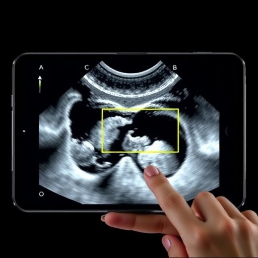
Ultrasound imaging has always been medicine’s great equalizer—cheap, safe, portable, and free of ionizing radiation. But it has also remained stubbornly flat. Where computed tomography and magnetic resonance imaging hand clinicians fully navigable three-dimensional volumes, a standard ultrasound exam delivers a stack of two-dimensional slices whose interpretation depends heavily on the skill of the person holding the probe. Now a research team reporting in the journal 3D Printing in Medicine has unveiled a deceptively simple system that converts a routine handheld ultrasound exam into a three-dimensional reconstruction using nothing more exotic than a printed QR code film stuck to the patient’s skin, a lightweight stereo camera clipped to the probe, and a clever localization algorithm borrowed from autonomous robotics. The work, led by Jiaojiao Ma and colleagues, promises to strip away one of the last major cost barriers separating everyday ultrasound from the spatial richness of 3D imaging.
The appeal of three-dimensional ultrasound is not merely cosmetic. Unlike a 2D sweep, a volumetric dataset captures the full geometry of an organ or lesion, exposing subtle abnormalities that can hide between standard scan planes and enabling precise quantitative analysis—volumes, surfaces, and spatial relationships computed directly from the image data. Such datasets also feed naturally into downstream technologies: virtual reality visualizations that help patients understand their own anatomy and engage more meaningfully with their care, and 3D printing pipelines that turn imaging data into physical surgical planning models. More recently, artificial intelligence and machine learning systems have begun exploiting 3D ultrasound data for automated segmentation and diagnosis, tasks that boost both efficiency and accuracy. Yet despite these advantages, the probes that generate 3D ultrasound have remained expensive, bulky, and constrained to a limited field of view. Creating an affordable, lightweight, wide-field 3D probe has become something of a holy grail for the field—one that demands progress in materials, manufacturing, transducer design, and software all at once.
The central obstacle in so-called freehand 3D ultrasound is knowing exactly where the probe is, and in which orientation, at the instant each 2D frame is captured. Only with that information can hundreds of tilted slices be stitched into a coherent volume. The gold-standard solutions—optical and electromagnetic trackers—do achieve real-time sub-millimeter accuracy, but they come with baggage. Optical tracking systems are costly and require an unobstructed line of sight between cameras and markers mounted on the probe. Electromagnetic trackers, while immune to line-of-sight issues, are notoriously susceptible to distortion from ferromagnetic metal in the environment, a serious problem in an operating theater full of steel instruments. Inertial sensing has occasionally been folded into cheaper schemes as well, but drift over time limits its standalone usefulness. Both mainstream approaches add hardware that many clinics, particularly in resource-limited settings, simply cannot justify. Earlier budget alternatives have included structured light to estimate probe orientation on the skin, optical-mouse-like sensors that track skin features over short distances, and strips of high-contrast markers laid along predefined scanning paths.
The new system takes a different route: it lets the patient’s own skin serve as the reference frame. Before scanning begins, a thin polypropylene film printed with a dense lattice of QR codes is smoothed onto the skin over the region of interest. The film is soft enough to conform to the body’s contours and, crucially, engineered so that it does not meaningfully attenuate the ultrasound beam passing through it into the tissue below. A compact stereo camera, rigidly bolted to the ultrasound probe by a custom 3D-printed bracket, watches this film continuously while the sonographer scans normally. Every camera frame captures a patch of the coded pattern, and the distinctive corner features of QR codes give the vision algorithms unmistakable landmarks for determining both position and orientation. Because the probe’s pose is computed relative to the patient’s body rather than the room, the approach is inherently robust to patient movement—an advantage over conventional external trackers.
Getting the film right required careful engineering trade-offs. The team settled on a code size of 1.4 millimeters per pixel after iterative testing: smaller codes choked the camera’s effective field of view and reduced feature density, while larger codes beyond roughly 2 millimeters sacrificed localization precision. The final design yields approximately 800 stable ORB features per frame—ORB, for oriented FAST and rotated BRIEF, being the fast corner-detection descriptor that underpins modern visual SLAM pipelines. The imaging hardware was tuned with equal deliberation. The camera, a binocular unit capable of recording at up to 1920 by 1080 resolution and 6 million pixels, was deliberately run at a modest 640 by 480 at 30 frames per second. Higher resolutions, the researchers found, overwhelmed their GPU’s processing capacity without improving tracking accuracy, while frame rates above 30 fps introduced data-handling latency that again bought no gains in reconstruction quality. A 2.3-centimeter focal length paired with a 5-centimeter working distance keeps the film squarely within the camera’s depth of field across the full 12-centimeter length of the probe.
The software pipeline then turns these synchronized streams of ultrasound frames and camera images into a navigable 3D model. Data from the Philips Lumify handheld probe and its attached camera travel over 5G or Wi-Fi to a backend server running Ubuntu with an NVIDIA RTX 3090 graphics card and Intel Xeon processor. There, the incoming images are preprocessed, a map of the coded skin surface—a “skin map”—is assembled from the accumulated camera frames, and an ORB-SLAM algorithm, the simultaneous localization and mapping technique popularized in autonomous vehicles and drones, analyzes the QR code features frame by frame to estimate the precise six-degree-of-freedom pose of the probe for every ultrasound image. A calibration step establishes the fixed geometric relationship between the camera’s field of view and the ultrasound image plane, so that each pixel in each slice can be assigned its true spatial coordinate. The positioned slices are then packaged into standard DICOM files, the universal language of medical imaging, and shipped to a volume-rendering platform built on the open-source 3D Slicer software, which assembles them into a three-dimensional model supporting multiplanar visualization and quantitative measurement.
To validate the system, the team put it through freehand scanning trials and model volume measurements. At a representative scanning speed of 15 millimeters per second, the ORB-SLAM positioning held its maximum cumulative error to no more than 2 percent across image acquisition frame rates of 10, 20, and 30 frames per second—performance the authors say satisfies the positioning requirements of their intelligent skin-texture image reconstruction system for thyroid ultrasound. The volume tests were arguably the more clinically meaningful yardstick: by reconstructing objects of known dimensions and comparing the volumes of the resulting 3D models against ground truth, the researchers confirmed that the stitched datasets are geometrically faithful, not merely visually plausible. The system successfully produced 3D reconstructions in which the features of a lesion and the spatial positions of the surrounding tissues could be displayed together in three-dimensional space—the kind of context that 2D imaging leaves the radiologist to mentally assemble slice by slice.
Thyroid imaging is a fitting proving ground. The gland sits close to the surface, within easy reach of a compact handheld probe, and nodular thyroid disease is among the most common reasons patients undergo ultrasound in the first place. Today, deciding whether a thyroid nodule warrants biopsy often hinges on how experienced the sonographer is at sweeping through the lesion and mentally reconstructing its shape, margins, and relationship to the trachea, vessels, and surrounding muscle. A volumetric reconstruction that lays out all of that geometry explicitly could shorten examinations and reduce operator dependence. And because the reconstructed volume can be re-examined in any plane long after the patient has left the clinic, suspected findings can be reviewed remotely by specialists without rescanning. The researchers envision the platform extending the same benefit across other superficial organs and eventually supporting image-guided interventions, where knowing the precise 3D position of a needle relative to a lesion is everything.
The broader significance lies in the economics. Handheld ultrasound probes that plug into tablets and smartphones have already distributed basic 2D imaging to remote clinics, ambulance crews, and low-resource hospitals worldwide; what they have lacked is a credible, affordable path to 3D. By replacing costly tracking hardware with a printed film, a commodity camera, and open-source reconstruction software, the new system points toward 3D ultrasound as a routine capability rather than a premium feature of flagship machines. Because the reconstruction runs on off-the-shelf hardware rather than bespoke electronics, the marginal cost of adding 3D capability to an existing handheld probe could be a tiny fraction of that of dedicated volumetric transducers. The platform is also deliberately standards-based, outputting DICOM files that plug into the existing ecosystem of PACS archives, AI analysis tools, and 3D printing workflows without proprietary lock-in—a design choice that lowers the barrier for other laboratories to build on the work.
Limitations remain, as the structure of the experiments makes clear. The film must be affixed smoothly before scanning, adding a small preparatory step to the clinical workflow, and the current validation centers on surface-accessible anatomy rather than deep abdominal organs. Accuracy at higher scanning speeds, performance across diverse skin properties, and long-term robustness in busy clinics will all require further study, though the researchers see room to grow through wider films, denser codes, and refined camera calibration. Still, the demonstration that commodity cameras, printed QR codes, and robotics-grade localization algorithms can meet the positioning tolerances of clinical 3D ultrasound marks a genuine inflection point. If follow-up studies replicate these error margins across more organ systems, the familiar grayscale sweep of an ultrasound exam could quietly become a full three-dimensional scan—in any clinic with a handheld probe, a tablet, and a sheet of coded film costing little more than a sticker.
Cite Scienmag News
Ophelia Keating. (August 30, 2026). Affordable handheld 3D ultrasound reconstruction system developed with visual positioning. Scienmag. https://scienmag.com/affordable-handheld-3d-ultrasound-reconstruction-system-developed-with-visual-positioning/
Ophelia Keating. "Affordable handheld 3D ultrasound reconstruction system developed with visual positioning." Scienmag, 30 August 2026, https://scienmag.com/affordable-handheld-3d-ultrasound-reconstruction-system-developed-with-visual-positioning/. Accessed 30 August 2026.
Ophelia Keating. "Affordable handheld 3D ultrasound reconstruction system developed with visual positioning." Scienmag. August 30, 2026. https://scienmag.com/affordable-handheld-3d-ultrasound-reconstruction-system-developed-with-visual-positioning/


