


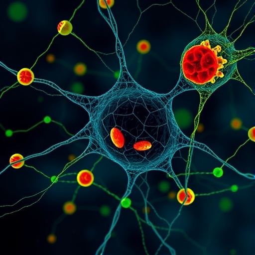
A new research paper introduces a method for watching metabolism at the level of individual cell types without relying on conventional physical labels, according to its title in Nature Methods. The technique, called REDCAT, is described as an “all-optical multimodal” system for mapping cell-type-specific metabolic activities. That combination of terms points to an effort to solve one of biology’s most persistent measurement problems: cells that look similar under a microscope can behave very differently, and their chemical activity can change rapidly in response to disease, treatment, nutrition or their surroundings. Rather than treating a tissue as a uniform mass, the approach is aimed at linking metabolic signals to particular kinds of cells while keeping the information spatially organized. The available source identifies the work as a 2026 research article by Y. Li, Z. Zhang, A. Enninful and colleagues, but does not provide experimental results, numerical performance data or a detailed description of the instrument. Its significance therefore lies in the measurement problem it targets and in the possibility of making cellular metabolism visible through optical readouts.
Metabolism is the network of chemical reactions that allows cells to obtain energy, build molecules and respond to stress. It includes processes such as glycolysis, in which glucose is broken down; oxidative phosphorylation, which produces much of a cell’s usable energy in mitochondria; lipid synthesis and degradation; amino-acid processing; and the production of chemical signals. These pathways are not distributed evenly across a tissue. A neuron, a muscle cell, an immune cell and a cancer cell may occupy the same microscopic environment while using different fuels and prioritizing different biochemical tasks. Even within one cell type, metabolic states can vary from one location to another. Traditional biochemical measurements often average signals across thousands or millions of cells, producing a powerful overall reading but concealing rare populations and local differences. A method that maps metabolic activity by cell type could therefore reveal patterns that disappear in bulk measurements, especially when a small group of cells drives inflammation, tissue repair or disease progression.
The phrase “single-cell-type-specific” is especially important because identifying individual cells is not the same as measuring their chemistry. Single-cell technologies can distinguish cells through gene-expression profiles, surface markers or physical shape, but a molecular identity does not automatically reveal what a cell is doing at a particular moment. Metabolism is dynamic: a cell can switch fuel sources, alter its energy production or redirect chemical building blocks within minutes or hours. A map that connects cell identity with metabolic activity would join two kinds of information that are often collected separately. It could show not only which cells are present, but also how those cells function in place. In a tissue, that distinction matters. Two neighboring cells might express similar markers yet experience different oxygen levels, nutrients or signals from nearby cells. Conversely, cells with different identities could converge on a common metabolic response during stress. REDCAT’s stated purpose is to capture these relationships using optical measurements.
“All-optical” suggests that the method is designed to acquire its information through light rather than requiring every molecular feature to be extracted by destructive biochemical processing. Optical approaches can use fluorescence, changes in light emission, absorbance, scattering or other light-sensitive signals to report on cellular conditions. Fluorescent sensors, for example, can be engineered or selected to respond to ions, redox state, enzyme activity or metabolites. When illuminated, they emit signals that can be recorded by a microscope or other detector. The advantage is that optical measurements can preserve spatial information and, in some cases, permit repeated observations of the same sample. The limitation is that light-based signals must be interpreted carefully. Fluorescence can vary with illumination intensity, sensor concentration, pH, oxygen availability and the optical properties of tissue. A signal may also represent a proxy for a metabolic process rather than a direct measurement of the molecule itself. Any useful system must therefore connect optical changes to defined biochemical states and control for technical sources of variation.
The word “multimodal” indicates that REDCAT is intended to combine more than one kind of optical information. In imaging, different modalities can provide complementary views: one signal may identify a cell, another may report energy production, and a third may indicate redox balance or the movement of a metabolic compound. Combining channels can be more informative than relying on a single color or measurement because metabolism is not one variable. A cell with high mitochondrial activity may also show altered redox chemistry, changes in lipid handling or a shift in the distribution of metabolites. Multimodal imaging can place these features into a common coordinate system, allowing researchers to ask whether they occur in the same cells and whether they are spatially linked. The approach also raises analytical challenges. Multiple signals must be calibrated, aligned and separated from background fluorescence. Computational models may be needed to classify cells, correct image distortion and distinguish biologically meaningful correlations from signals that merely change together because of the imaging conditions.
A central attraction of optical mapping is its ability to retain context. In a conventional metabolic assay, tissue can be homogenized and analyzed with mass spectrometry, enzymatic tests or other chemical methods. Those techniques can be exceptionally sensitive and can identify many compounds, but homogenization removes the original locations of the molecules and mixes signals from different cell populations. Imaging reverses that trade-off: it preserves geography, showing where activity occurs, but often measures a narrower set of signals and requires careful validation. A cell’s position can be biologically decisive. Metabolic behavior may differ near a blood vessel, at the edge of a tumor, beside an inflamed region or within a specialized tissue compartment. Mapping can expose gradients and neighborhoods that bulk assays cannot resolve. If REDCAT achieves the scope implied by its title, it could provide a framework for examining these patterns while simultaneously assigning them to specific cell types.
The potential applications are broad, although the supplied information does not establish that the method has already been applied successfully to any particular disease or biological system. In cancer research, metabolic maps could help distinguish tumor cells from immune and stromal cells and reveal how their chemical programs interact. In immunology, they could clarify whether activated immune cells occupy distinct metabolic niches or compete with neighboring cells for nutrients. In neuroscience, spatial measurements might help connect neuronal activity with the energy use of support cells. In developmental biology, they could track how metabolic programs change as cells specialize. The same logic could be relevant to drug development: a treatment might suppress a disease-associated metabolic state in one cell population while producing an unintended response in another. Cell-type-resolved measurements could make such effects easier to detect. These are prospective uses, not findings reported in the limited source material, and they would depend on the method’s sensitivity, specificity, speed and compatibility with living or preserved samples.
The technical standard for such a platform will be high. To claim cell-type-specific metabolic activity, researchers must establish how cell types are recognized and how reliably the optical signals correspond to metabolism. A convincing analysis would need controls showing that the signals change when a pathway is chemically or genetically perturbed, and that the method can distinguish genuine metabolic differences from variations in cell size, illumination or sensor expression. Spatial resolution is another critical factor: if neighboring cells are smaller than the microscope’s effective resolution, their signals may blur together. Temporal resolution determines whether the technique can follow rapid metabolic transitions or only capture stable states. Multiplexing introduces additional problems because fluorescent channels can overlap, while intense illumination can damage cells or bleach reporters. The title alone does not reveal how REDCAT addresses these issues, but naming the method as an all-optical multimodal mapping strategy places it within a rapidly developing effort to make cellular chemistry measurable in its native spatial setting.
What makes the work potentially newsworthy is the prospect of turning metabolism from an averaged laboratory measurement into a visually organized property of living systems. Biology increasingly depends on understanding interactions among cell types rather than studying each population in isolation. A tissue is not merely a collection of cells; it is a chemical landscape in which cells exchange nutrients, signals and waste products. Tools that connect identity, location and function could help researchers see that landscape with greater precision. REDCAT’s reported focus is not simply on taking more images, but on integrating optical information into maps that distinguish metabolic activity among specific cell types. The source provided for this report contains only the article’s bibliographic record and title, so claims about performance, discoveries or biological conclusions cannot be independently assessed here. Even so, the approach reflects a major direction in modern cell biology: measuring what cells are doing, where they are doing it and how their chemistry changes in relation to their neighbors. If validated across biological settings, such technology could make hidden metabolic differences visible—and give researchers a sharper view of how tissues function in health and disease.
Cite Scienmag News
Violet A. (August 28, 2026). REDCAT Enables All-Optical Multimodal Mapping of Metabolism in Specific Cell Types. Scienmag. https://scienmag.com/redcat-enables-all-optical-multimodal-mapping-of-metabolism-in-specific-cell-types/
Violet A. "REDCAT Enables All-Optical Multimodal Mapping of Metabolism in Specific Cell Types." Scienmag, 28 August 2026, https://scienmag.com/redcat-enables-all-optical-multimodal-mapping-of-metabolism-in-specific-cell-types/. Accessed 28 August 2026.
Violet A. "REDCAT Enables All-Optical Multimodal Mapping of Metabolism in Specific Cell Types." Scienmag. August 28, 2026. https://scienmag.com/redcat-enables-all-optical-multimodal-mapping-of-metabolism-in-specific-cell-types/


