

A tiny implant designed to wedge open one of the most delicate joints in the human spine has passed a series of demanding laboratory tests—and, in a patient-specific 3D-printed model, cut the force needed to realign the upper neck by about 70 percent. The device, an expandable titanium-alloy spacer developed for difficult cases of atlantoaxial subluxation, is intended to help surgeons correct a deformity that can leave the spinal cord compressed and, in severe cases, cause permanent neurological damage. The study, published in 3D Printing in Medicine, combines implant engineering with medical imaging and additive manufacturing to investigate whether a new mechanical approach could make reduction of the C1–C2 joint more controlled and less forceful.
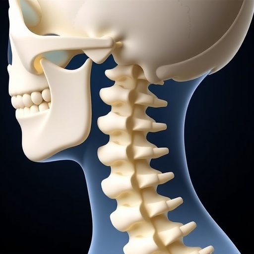
The atlantoaxial joint is formed by the first two cervical vertebrae, known as the atlas, or C1, and the axis, or C2. It is responsible for much of the head’s rotation while also surrounding and protecting the upper spinal cord. Injury, rheumatoid arthritis, degeneration, congenital abnormalities, tumors, or changes in the odontoid process—the tooth-like projection of C2—can destabilize the joint. In atlantoaxial subluxation, C1 shifts relative to C2. Patients may initially experience neck pain, but progressive displacement can narrow the space available for the spinal cord, producing compressive myelopathy, weakness, loss of coordination, or paralysis.
Realigning the joint is especially challenging when the atlas has tilted forward and the articular surfaces have become locked together by bone growth, osteophytes, scar tissue, or ankylosis. Conventional posterior traction or screw-and-rod systems can stabilize the neck, but they may not fully correct a fixed deformity. Pulling directly on C1 in such cases can force the vertebra along the wrong path: instead of moving toward its normal position, it may slide parallel to the inclined joint surfaces. That inefficient trajectory can demand greater traction, increase stress on surrounding ligaments and soft tissues, and leave residual spinal-cord compression. Joint release and interfacet distraction are already used to address some of these problems, but most existing spacers are static and generally have limited ability to correct the forward inclination of the atlas.
The new implant was designed as a tapered wedge, with a 2-millimeter anterior tip and a 10-millimeter posterior height, producing an angle of roughly 17 degrees. Its contact surface measures 18 millimeters long by 10 millimeters wide, dimensions selected using CT measurements from 144 people with anatomically normal atlantoaxial joints. Serrated teeth on its upper and lower surfaces are intended to grip the opposing facet surfaces and resist migration. Unlike a conventional fixed cage, the spacer contains a staged internal mechanism. A posteriorly inserted screw first expands the anterior part of the implant, opening the front of the joint and lifting the atlas. An inner rod then advances through the screw and pushes a central serrated blade outward, increasing contact pressure and fixation stability.
The sequence is intended to change the geometry of the joint before posterior traction is applied. By raising the front of C1, the spacer reduces the abnormal forward inclination and redirects the force used for reduction toward the physiological axis of the spine. A custom handle was developed to perform the procedure in stages. The instrument holds the spacer during insertion, activates the first expansion by rotating a control ring, deploys the central blade with a push rod, and connects to a C1 screw through a side rod. In principle, this allows distraction of the joint and posterior traction on the atlas to occur as a coordinated maneuver rather than as separate, competing forces.
The researchers tested the implant’s structural performance before examining its reduction behavior. Three devices underwent static axial compression, producing an average yield load of 6,725 ± 123 newtons. That result exceeded the 5,450-newton minimum cited in ISO 23089-2 for cervical fusion devices and was above the approximate 4,000–6,000-newton range reported for some commercially available implants. In fatigue testing, the spacers endured more than 5 million loading cycles without visible cracking, deformation, or component failure. The cyclic force ranged from 165 to 1,650 newtons, with the upper value set at the recommended cervical-device runout load of 1,500 newtons multiplied by a safety factor of 1.1. The findings suggest that the mechanism can withstand repeated compression in laboratory conditions, although they do not by themselves establish how the implant would perform in living bone.
The expansion mechanism also appeared to be mechanically manageable. Under a constant 5-kilogram-force load, the researchers rotated the internal screw through three full turns while recording torque. Full deployment required an average maximum torque of 52.56 ± 7.54 newton-millimeters. That value is small compared with the torque commonly associated with pedicle-screw insertion or expandable lumbar cages, and it indicates that the device could potentially be deployed with controlled manual force. Keeping expansion torque low matters in the upper cervical spine, where a surgeon must manipulate a compact implant through a narrow joint while avoiding uncontrolled movement, jamming, or damage to the surrounding anatomy.
To test reduction, the team reconstructed a severely deformed C1–C2 joint from high-resolution clinical CT data, with slices 0.625 millimeters thick. The digital images were segmented and refined while preserving the patient’s anterior inclination, osteophytes, and degenerative joint shape. The resulting models were printed from ABS polymer using fused-deposition modeling, with 0.254-millimeter layers and 80 percent infill. The researchers oriented the vertebrae vertically to reduce stair-step artifacts on the joint surfaces, then smoothed the models with acetone vapor and checked their dimensions to within 0.2 millimeters. Springs placed between C1 and C2 represented ligamentous resistance, with a calibrated stiffness of 0.4 newtons per millimeter. The atlantodental interval, the gap between the atlas and the odontoid process, was set at 10 millimeters to reproduce the dislocated configuration.
The model allowed the investigators to compare traction with and without the expandable spacer under otherwise identical conditions. Steel cables attached to C1 were pulled upward at 5 millimeters per minute until the atlantodental interval closed. Without the spacer, the maximum reduction force was approximately 111.7 ± 5.7 newtons. The atlas initially resisted movement as its articular surfaces remained mechanically locked, then shifted as the surfaces disengaged; motion tracking showed that C1 traveled mainly parallel to the joint surface rather than along the applied traction vector. With the spacer inserted, the maximum force fell to about 35.3 ± 3.2 newtons—roughly a 68 percent reduction—and the movement path became smoother and more closely aligned with the intended vertical direction. The smaller displacement amplitudes recorded at tracking landmarks also suggested that reduction occurred with less deformation of the model’s simulated soft tissues.
Those results make the implant an intriguing candidate for difficult atlantoaxial reductions, but they remain preclinical evidence rather than proof of clinical benefit. The study used only three specimens for each mechanical test and a single patient-derived anatomy for the reduction platform. Printed polymer does not reproduce the complex behavior of cortical bone, cancellous bone, cartilage, capsules, muscles, and ligaments, and springs cannot capture all the nonlinear, three-dimensional constraints present in a living neck. The authors also note that the implant dimensions were developed around anatomical data from an Asian population and may require adaptation for other groups. The final spacer was manufactured by precision CNC machining rather than metal 3D printing because the multi-component sliding and threaded mechanism demands tight tolerances and reliable surface quality. Even so, the 3D-printed model offers a powerful bridge between design and surgery: it can reproduce patient-specific deformity, make ligament tension adjustable, and provide repeatable measurements when suitable cadaveric specimens are scarce. Further testing in broader anatomical models, cadaveric tissue, and eventually carefully controlled clinical studies will be needed to determine whether the promising 70-percent reduction in laboratory traction translates into safer real-world treatment for patients facing one of the spine’s most technically demanding operations.


